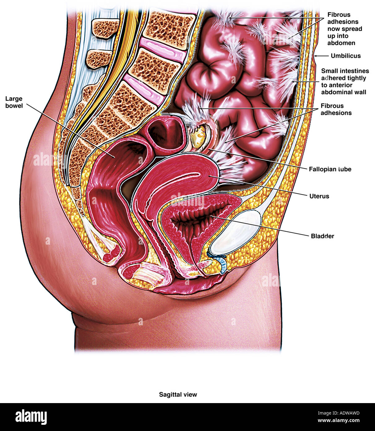
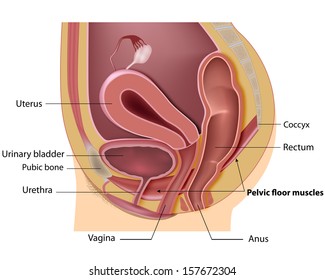
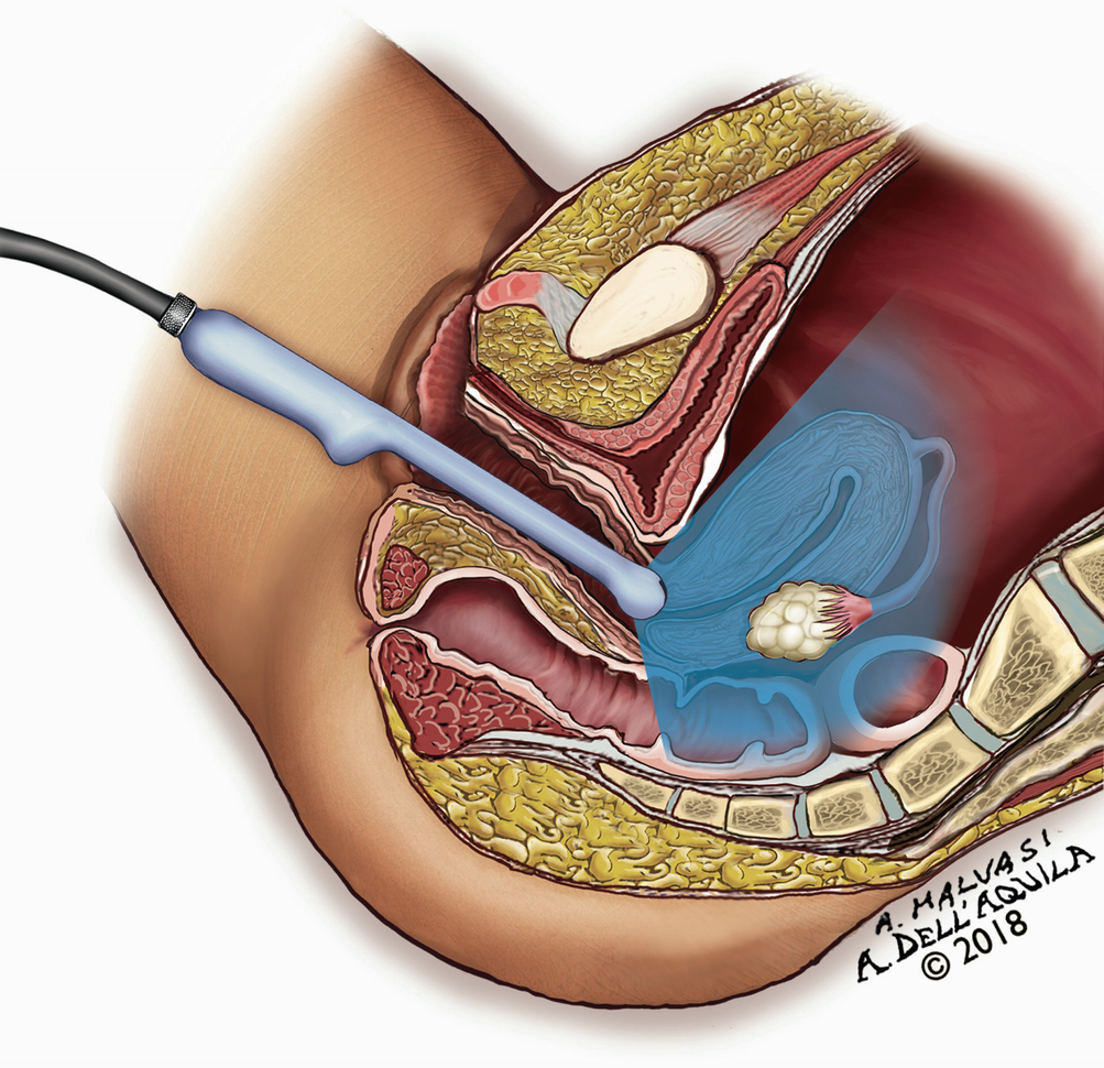
Anatomy of the female pelvis. A sagittal view of the female pelvis is shown in the figure.
Female Pelvic Anatomy
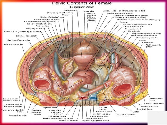
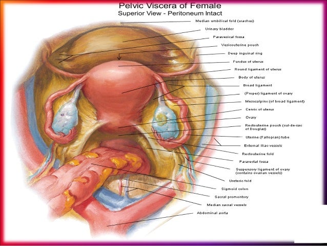
The female pelvic organs include the egg producing ovaries and the uterine tubes that carry the eggs into the uterus for potential fertilization by male sperm.

Female pelvic anatomy. This is a patient education video on normal female pelvic anatomy. The wider inlet facilitates head engagement and parturition. What is the female pelvis.
They also include the vagina which is the entryway to the uterus. Using a speculum a doctor can examine the vulva vagina and cervix. This area provides support for the intestines and also contains the bladder and.
The anatomy of the lower genital tract comprised of the vulva and vagina is discussed separately. The strength of the pelvic muscles can also be tested. The female pelvis figure 1a has a wider diameter and a more circular shape than that of the male.
The female upper genital tract consists of the cervix uterine corpus fallopian tubes and ovaries. The male inlet is more heart shaped. The female pelvis is larger and broader than the male pelvis which is taller narrower and more compact.
The sides of the male pelvis converge from the inlet to the outlet whereas the sides of the female pelvis are wider apart. Its located between the abdomen and the legs. Axial slice showing uterus ovary uterine tubes ligament vaginal cavity and other internal organs.
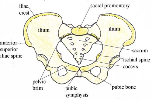
Anatomy of the human female pelvis. Bony pelvis and pelvic joints sacrum coccyx and innominate bones ilium ischium and pubis. The strength of the pelvic muscles can also be tested.
The pelvis is the lower part of the torso. How to use the anatomical labels. The inferior pelvic outlet is closed by the pelvic floor.
This is a patient education video on normal female pelvic anatomy. The female inlet is larger and oval in shape while the male sacral promontory projects further ie. Fuse at acetabulum ilium articulates with the sacrum posteriorly at sacroiliac joint synovial joint stability of the bony pelvis pubic bones articulate with each other anteriorly at symphysis pubis cartilaginous joint.
See surgical female urogenital anatomy section on lower genital tract.
 Abdominal And Pelvic Anatomy Female Stock Photo 7713052
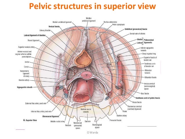 2 Female Pelvic Anatomy Warda Part 2
 3b Scientific H20 3 Female Pelvis W Ligaments 4 Part 3b Smart Anatomy
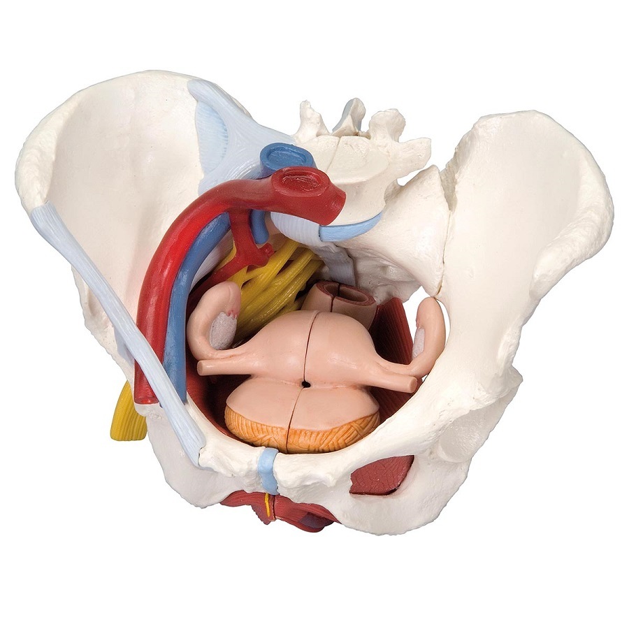 Anatomical Models Of Female Pelvis With Ligaments Vessels
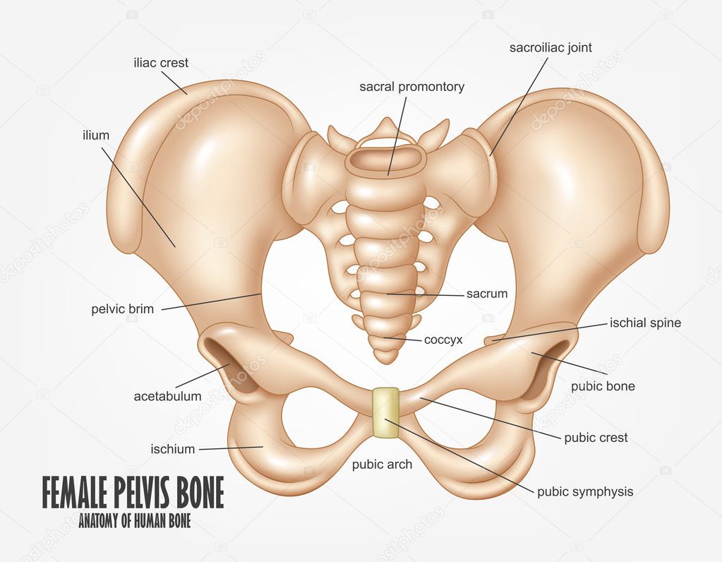 Diagram Of Female Pelvic Bone Female Pelvis Bone Anatomy
5 Anatomical Detail Of Female Pelvic Anatomy Adapted From
 Female Pelvic Applied Anatomy By Dr Shashwat Jani
 Female Pelvic Anatomy Stack Print Lyon Road Art
 Human Animal Anatomy And Physiology Diagrams Normal Anatomy
 Drawing Of Female Pelvis Midsagittal View Shows The
 Female Anatomy The Functions Of The Female Organs Hers
 Female Pelvic Anatomy
 Antenatal Care Module 6 Anatomy Of The Female Pelvis And
 The Ultimate Pelvic Anatomy Resource Pelvic Guru Featured
 Female Pelvic Applied Anatomy By Dr Shashwat Jani
 Female Pelvic Anatomy Medical Illustration Medivisuals
 The Ultimate Pelvic Anatomy Resource Pelvic Guru Featured
 Pelvic Anatomy Obgyn Net
 Female Pelvis Model With Ligaments Vessels Nerves Pelvic Floor And Organs Life Size 6 Part
 Imagenes Fotos De Stock Y Vectores Sobre Female Pelvic
Female Pelvis Cross Section Model
 Female Pelvic Anatomy Diagram Quizlet
 Pelvic Floor Muscles The Facts Continence Foundation Of
 Normal Ultrasound Female Pelvic Anatomy Springerlink


Posting Komentar
Posting Komentar