Venous system overview vein structure. Anatomy of the venous system.

Venous return vr is the volume of blood that reaches the right heart.
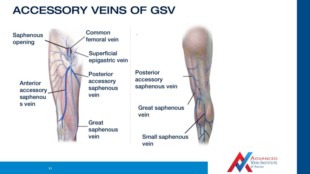
Venous anatomy. The superficial subcutaneous venous system in the legs includes the long saphenous vein and the short saphenous vein. Treatment including angioplasty and stent placement can open the vein but it tends to narrow again restenosis. This is the outer layer of the vein wall.
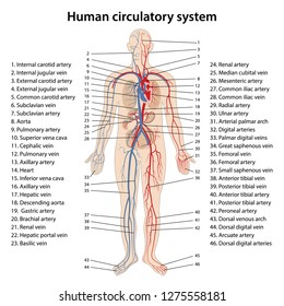
Veins are the blood vessels which carry the blood from peripheral tissues towards heart. We distinguish between the superficial and the deep venous systems. Sometimes vein problems can occur most commonly due to either a blood clot or a vein defect.
Venous drainage anatomy overview. They carry the deoxygenated blood which is bluish in color and for the same reason veins appear blue. Effective communication and an understanding of the treatment options require a common nomenclature as updated by an international consensus committee.
Pulmonary vein stenosis is a condition in which the pulmonary vein is thickened leading to narrowing. Varicose veins without skin changes are present in about 20 of the general population and they are slightly more frequent in women. Venous anatomy is divided into three systems.
Veins have thinner walls and larger lumina than arteries do. Veins are less muscular than arteries and are often closer to the skin. A primary characteristic of the deep veins is that they run alongside the arteries and as such often share the same name.
It is an uncommon but serious birth defect and is often combined with other heart abnormalities. The anatomy of the lower extremity venous system is complex and highly variable. Unlike the high pressure arterial system the venous system is a low pressure system that relies on muscle contractions to return blood to the heart.
Grossly the venous system is composed of venules and small and great veins. Exceptions are the pulmonary and umbilical veins both of which carry oxygenated blood to the heart. Use this interactive 3 d diagram to explore the venous system.
Chronic venous diseases cvds include a spectrum of clinical findings ranging from spider telangiectasias and varicose veins to debilitating venous ulceration. The venous system is that part of the circulation in which the blood is transported from the periphery back to the heart. Deep superficial and perforating.
In contrast to veins arteries carry blood away from the heart. Veins are often categorized based on their location and any unique features. Most veins carry deoxygenated blood from the tissues back to the heart.
Tips for healthy.

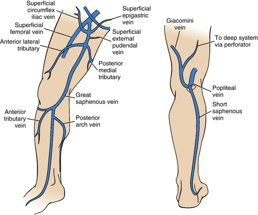
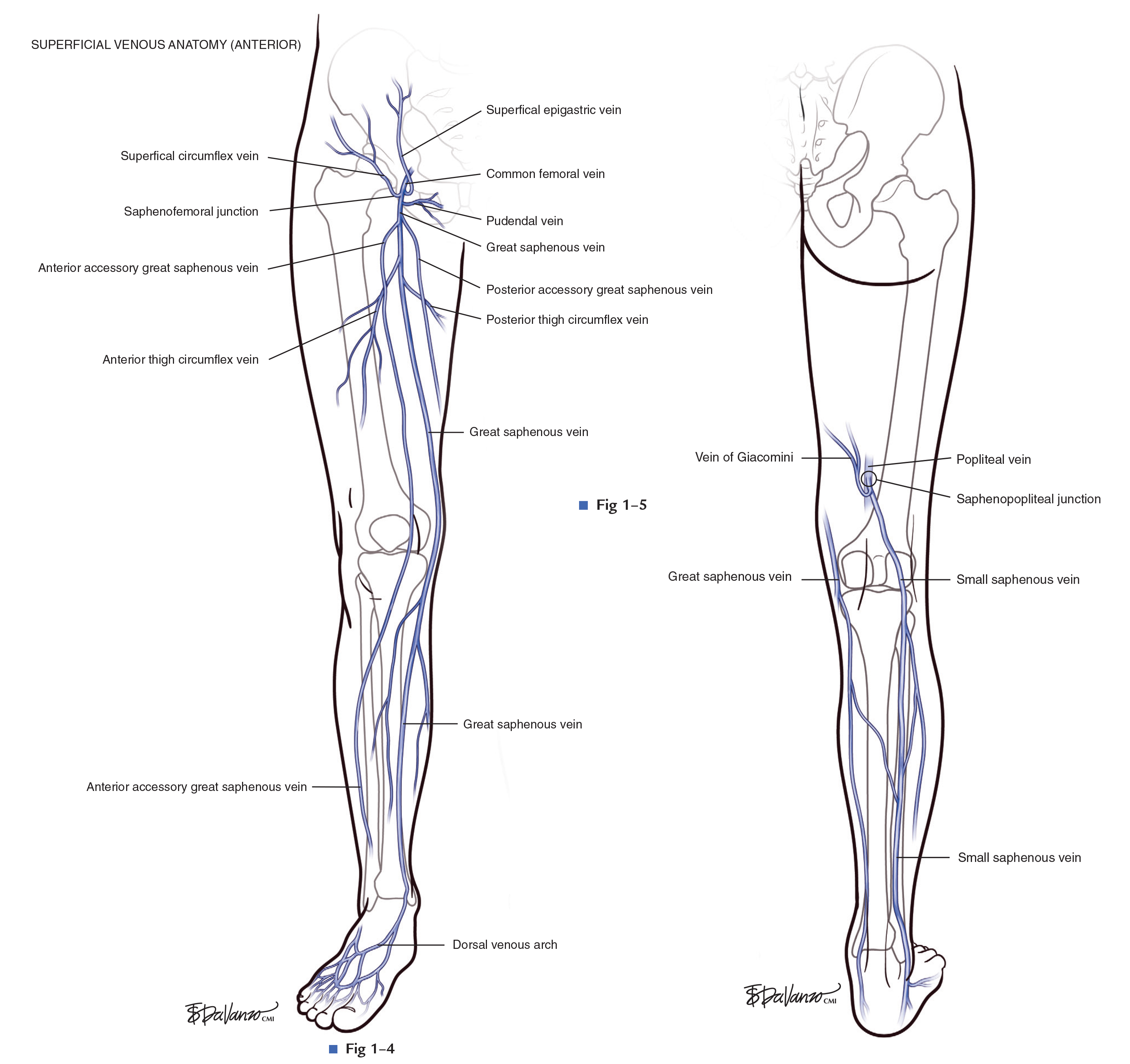
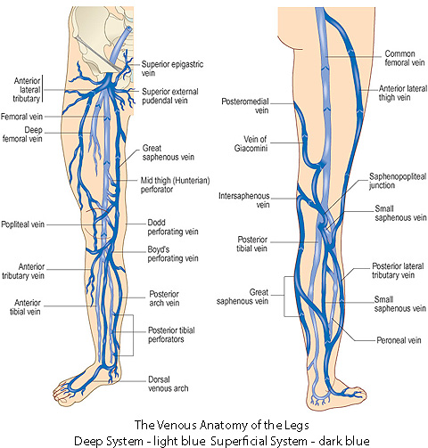
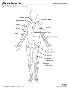
 Lower Extremity Venous Anatomy Dallas Tx Venous System
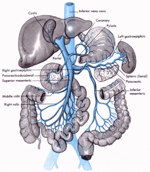
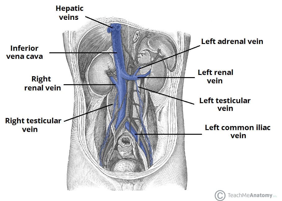 Venous Drainage Of The Abdomen Teachmeanatomy
 Venous Insufficiency Redbacteria
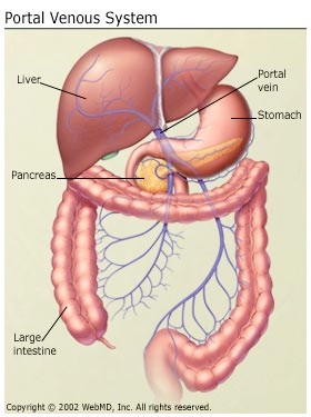
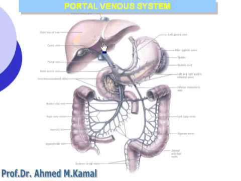 1 Portal Venous System
 Venous System Stock Vectors Images Vector Art Shutterstock
 Veins Types Venous System Clinical Significance How To
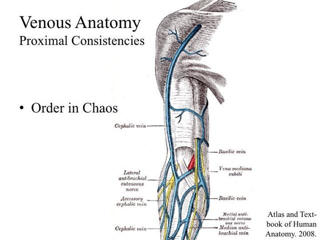
 Ecr 2013 C 2631 Upper Extremity Venous Ultrasound


 Vein Wikipedia
 Schematic View Of Venous Anatomy From Insightful Phlebology

 Venous System
 Science Source Venous System Of The Pelvis Artwork
 Venous Drainage Anatomy Overview Microscopic Anatomy
 J Neurosurgery On Twitter Neurosurgicalatlas
 Human Venous System Diagram Quizlet
 Which Three Venous Systems Are Affected By Chronic Venous
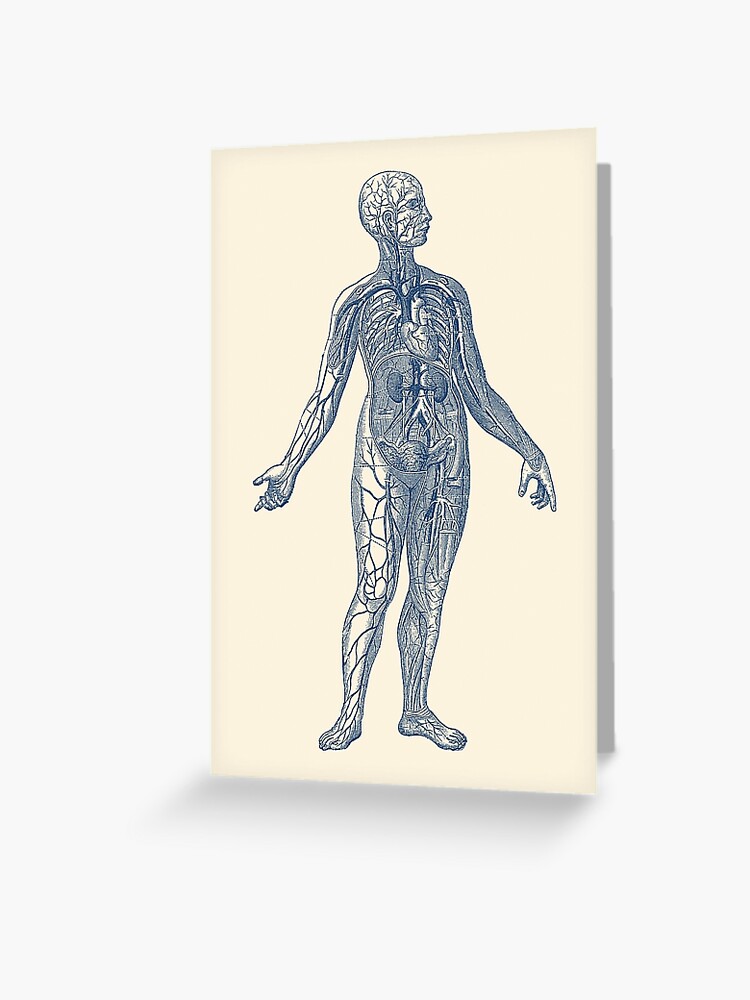 Venous System Diagram Vintage Anatomy Greeting Card
Lower Extremity Veins Radiology Key
 Video The Venous Anatomy Varicose Vein Treatment In Tempe
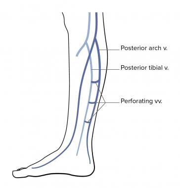
 Diagram Showing The Venous Anatomy Of The Leg
 Abdomen Venous Portal System Ranzcrpart1 Wiki Fandom


Posting Komentar
Posting Komentar