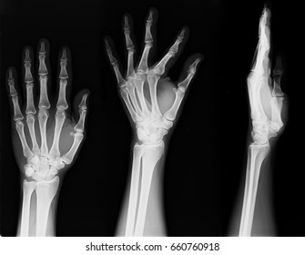
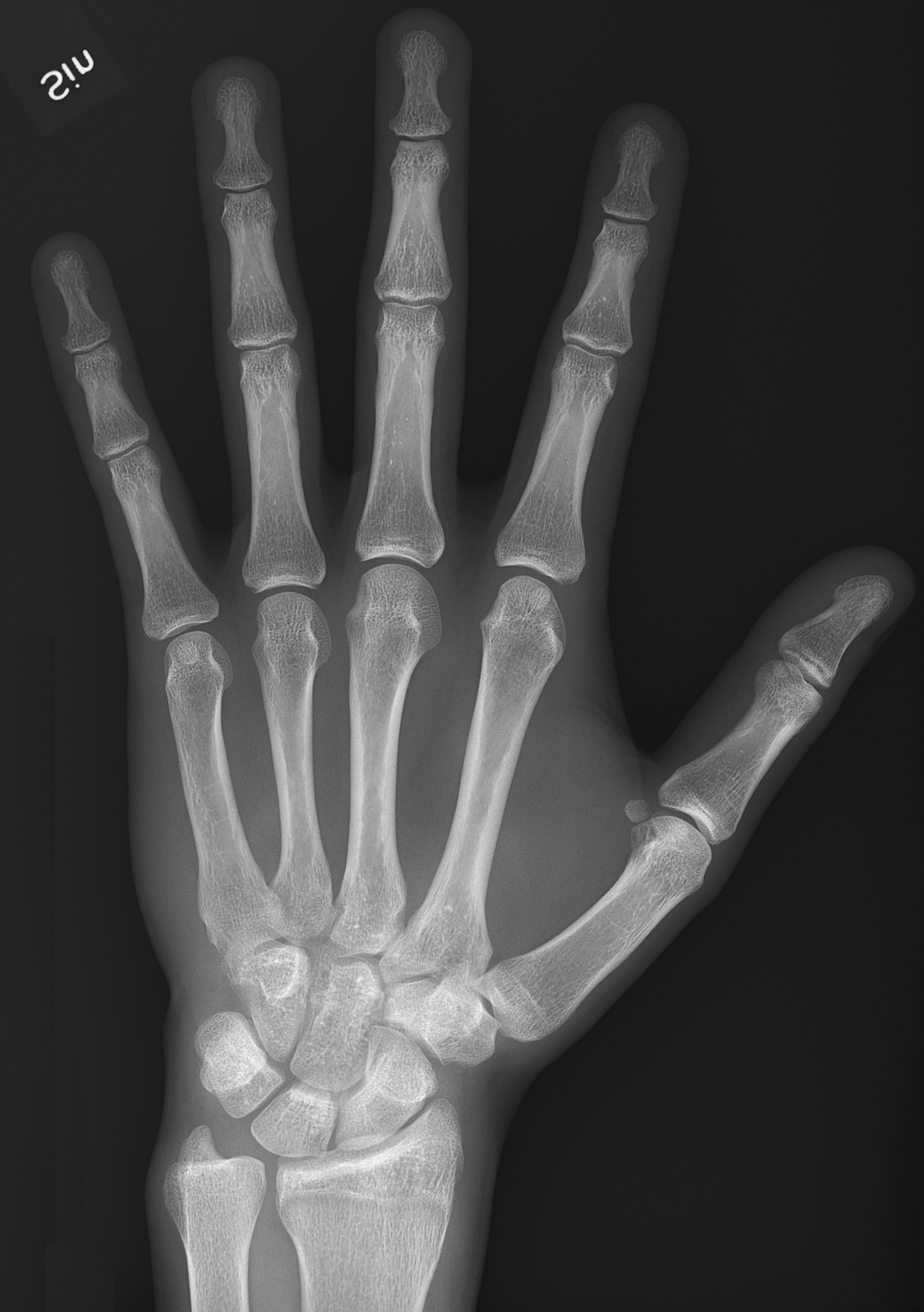
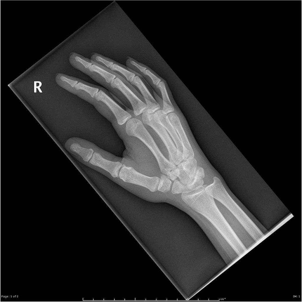
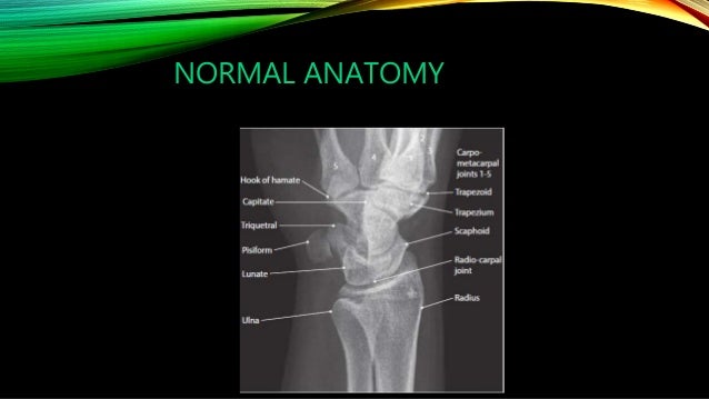
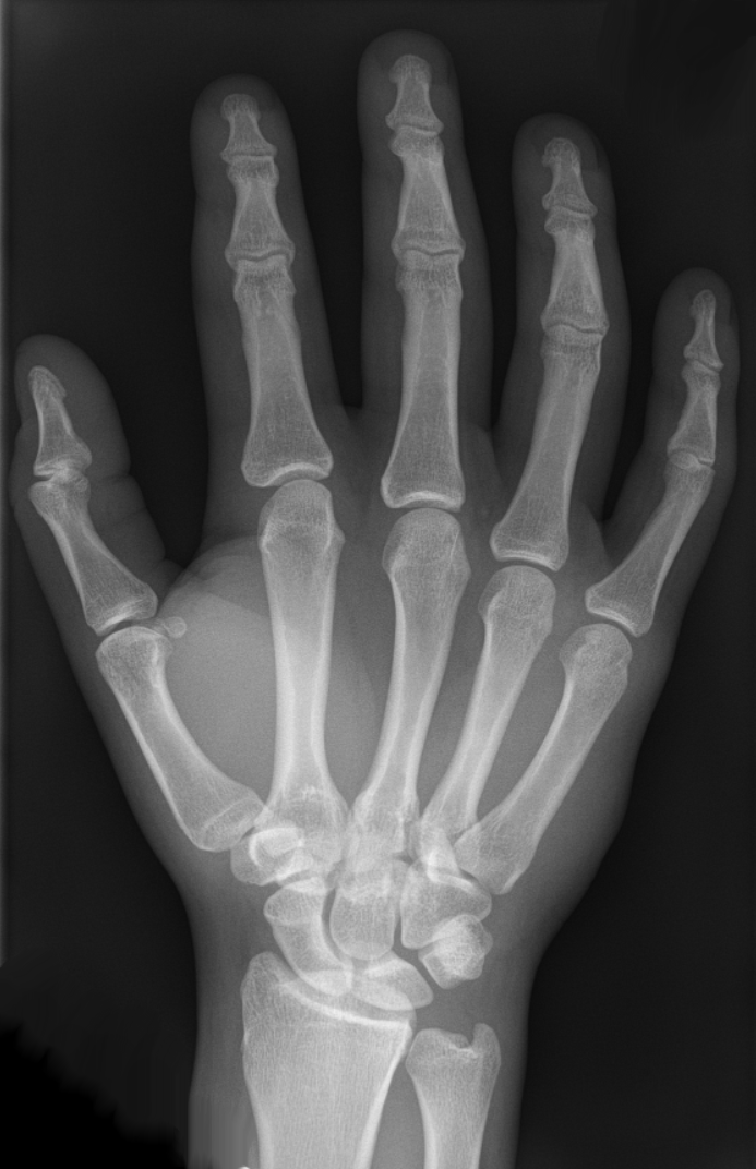
A hand radiograph contains a pa and oblique view of the distal radius and ulna and the carpus. The series primarily examines the radiocarpal and distal radioulnar joints the carpals metacarpals and phalanges.
Case contributed by dr benoudina samir.

Hand anatomy xray. For right now we gather some pictures of hand anatomy bones xray and each of them displaying some new inspiration. Adult hand radiographic anatomy. Play this quiz called hand xray anatomy and show off your skills.
Adjustments in kvp and mas should be considered in cases involving splints casts wraps swelling braces etc. 3 article feature images from this case. The joint spaces of the cmc joints are equal average 1 2 mm and form a zigzag configuration fig.
A basic review should start with ap and lateral views including the entire foot and ankle. Login register free help. After showing this hand anatomy bones xray i can guarantee to aspire you.
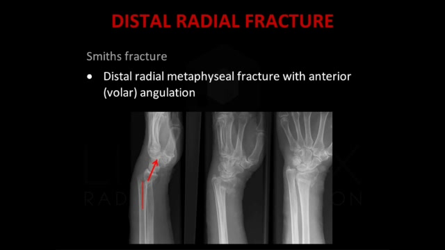
Check the wrist as you would for a wrist radiograph an approach distal radius. Although additional radiographs can be taken for specific indications. Finger injuries visible on x ray include bone fractures dislocations and avulsions the hand comprises the metacarpal and phalangeal bones.
Symmetrical joints where the bones do not overlap except the carpal bones and the base of the metacarpal bones. Hand radiograph an approach hand series. One of the commonest misses in trauma films of the hand and wrist is a dislocation of the 5th carpometacarpal joint which may cause significant morbidity if the diagnosis is delayed.
Normal radiographic anatomy of the hand. Adult hand radiographic anatomy. Wrist radiograph an approach.
Anatomy hand identify phalanges xray hand xray hand anatomy pa hand oblique hand identify anatomy. This is a quiz called hand xray anatomy and was created by member desimichelle. Anatomy of the lateral wrist figure 2.
This article relates mainly to the traumatic injuries to the foot. Diagnosis certain diagnosis certain. Other games by same author.
Anatomy of the lateral wrist. Hand x rays are indicated for a variety of settings including. Often a foot x ray is also requested for the investigation of osteomyelitis arthritides or a bone lesion.
Characteristics of a normal handfinger x ray. The hand anatomy bones xray could be your choice when creating about bone. Fractures and dislocations are usually straightforward to identify so long as the potentially injured bone is fully visible in 2 planes.
Although x ray machines vary the general kvp ranges for radiography of the wrist and hand is between 50 65 kvp. Drag here to reorder. The hand series consists of a posteroanterior oblique and a lateral projection.
Keep the body part as close to the cassette as possible in order to reduce oid object image distance.
 Wrist Xray Images Stock Photos Vectors Shutterstock
Normal Pediatric Bone Xrays Bonexray Com
 File X Ray Of Normal Hand By Dorsoplantar Projection Jpg
Uams Gross Anatomy X Ray Atlas
 Scientists Are Putting The X Factor Back In X Rays Popular
Bone Age A Handy Tool For Pediatric Providers

Hand Radiographic Anatomy Wikiradiography
 Amazon Com Ahawoso Canvas Prints Wall Art 16x16 Inches
 Nursing X Ray Interpretation Part 7 Wrist And Hand
 The Radiology Assistant Wrist Carpal Instability
 How To Read A Hand Wrist Xray For Orthodontics Straightsmile Solutions Method
 Infographic Diagram Of Human Hand Bone Anatomy System Anterior
 Normal Hand Radiographs Radiology Case Radiopaedia Org
 Papercut No 6 Skeleton Hand Xray By Nicky Krusch On
 Carpal Ossification Radiology Case Radiopaedia Org
 Basilar Thumb Arthritis Hand Orthobullets
Standardised Post Operative Radiographs For Volar Radial


Posting Komentar
Posting Komentar