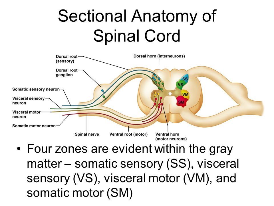
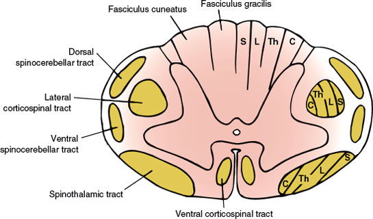
Cross sectional anatomy of the spinal cord. Grey matter consists of neuron cell bodies with little myelin and white matter consists of myelinated axons.
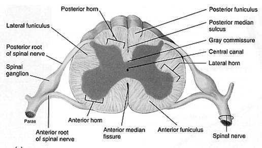 Cross Sectional Anatomy The Central Nervous System
It is covered by the three membranes of the cns ie the dura mater arachnoid and the innermost pia mater.

Sectional anatomy of the spinal cord. The spinal cord finishes growing at the age of 4 while the vertebral column finishes growing at age 14 18. Almost dividing the spinal cord in half are two longitudinal grooves. Two prominent grooves or sulci run along its length.
The spinal cord like the brain consists of two kinds of nervous tissue called gray and white matter. Describe the cross sectional anatomy of the spinal cord. Gray matter has a relatively dull color because it contains little myelin.
The spinal cord is part of the central nervous system cns. Spinal cord segments cross sectional anatomy. It is situated inside the vertebral canal of the vertebral column.
Central area of grey matter shaped like a butterfly and surrounded by white matter in three columns. Cross sectional anatomy of spinal cord. It passes through the spinal canal or spinal cavity of the vertebral column ie the backbone or spine.
An interactive quiz covering spinal cord cross sectional anatomy through multiple choice questions and featuring the iconic gbs illustrations. Contains 2 types of nervous tissue. The gray matte of the spinal cord is located at its center with the white matter surrounding it.
Gross anatomy the spinal cord is part of the central nervous system cns which extends caudally and is protected by the bony structures of the vertebral column. Spinal cord anatomy basically spinal cord is a long and narrow bundle of nervous tissues and support cells which extends from the base of our brain to the upper lumbar region. The two grooves are named as follows.
The central gray matter contains the neural cell bodies. It contains the somas dendrites and proximal parts of the axons of neurons. The spinal cord is elliptical in cross section being compressed dorsolaterally.
The ventral anterior median fissure and the more shallow dorsal posterior median sulcus. These two grooves run the length of the cord and partially divide it into right and left halves. The narrow indentation that partitions the back surface is known as the dorsal median sulcus or posterior median sulcus and the broader groove that partitions the front surface is called the ventral median fissure or anterior median fissure.
The posterior median sulcus is the groove in the dorsal side and the anterior median fissure is the groove in the ventral side. The peripheral white matter contains the axon tracts. A cross sectional view of the spinal cord demonstrates a central butterfly shaped area of gray matter and peripheral white matter fig.
Tracts are named with their point of origin first. During development theres a disproportion between spinal cord growth and vertebral column growth.
Cross Sectional Anatomy The Central Nervous System
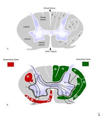 Lower Cervical Spine Fractures And Dislocations Background
 Biology Pictures Spinal Cord Crossection Spinal Nerves
 The Spinal Cord Human Anatomy And Physiology Lab Bsb 141
 Cns Cross Sectional Anatomy Of Spinal Cord Flashcards
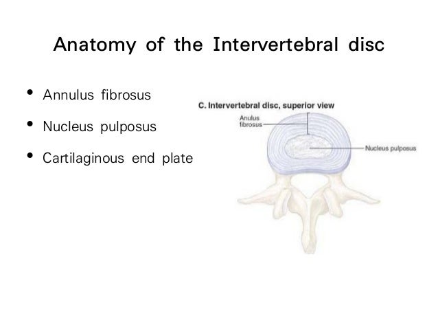 Applied Cross Sectional Anatomy Of Spinal Cord
 Spinal Cord Anatomy Parts And Spinal Cord Functions
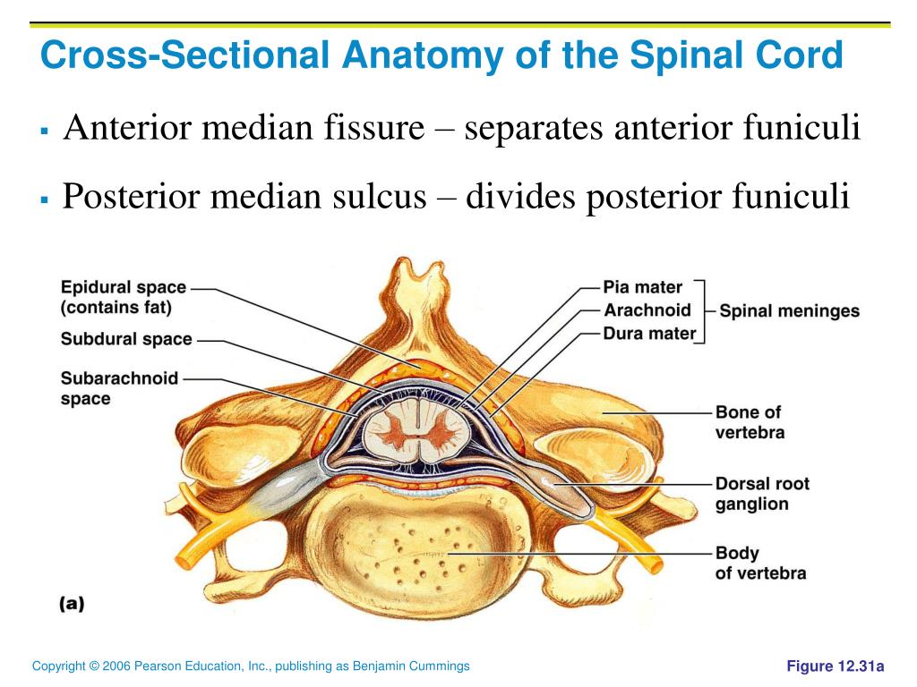 Ppt The Central Nervous System Powerpoint Presentation
Spinal Cord Ventral Lateral Surface Cross Sectional Quiz
 Solved Or Review Practice Sheet Exercise Ben Eay 312011
 Chapter 12b Spinal Cord Ppt Video Online Download
 Applied Cross Sectional Anatomy Of Spinal Cord
 Lecture 14 Chapter 13 Spinal Cord Ppt Download
 General Cross Sectional Anatomy Of The Spinal Cord
Ch 12 Gross Anatomy Of The Spinal Cord
 Sectional Anatomy Of A Spinal Column Purposegames
 Applied Cross Sectional Anatomy Of Spinal Cord
 Applied Cross Sectional Anatomy Of Spinal Cord
 Applied Cross Sectional Anatomy Of Spinal Cord
 Pocket Atlas Of Sectional Anatomy Volume 1 Pages 251 272
 Duke Neurosciences Lab 2 Spinal Cord Brainstem Surface
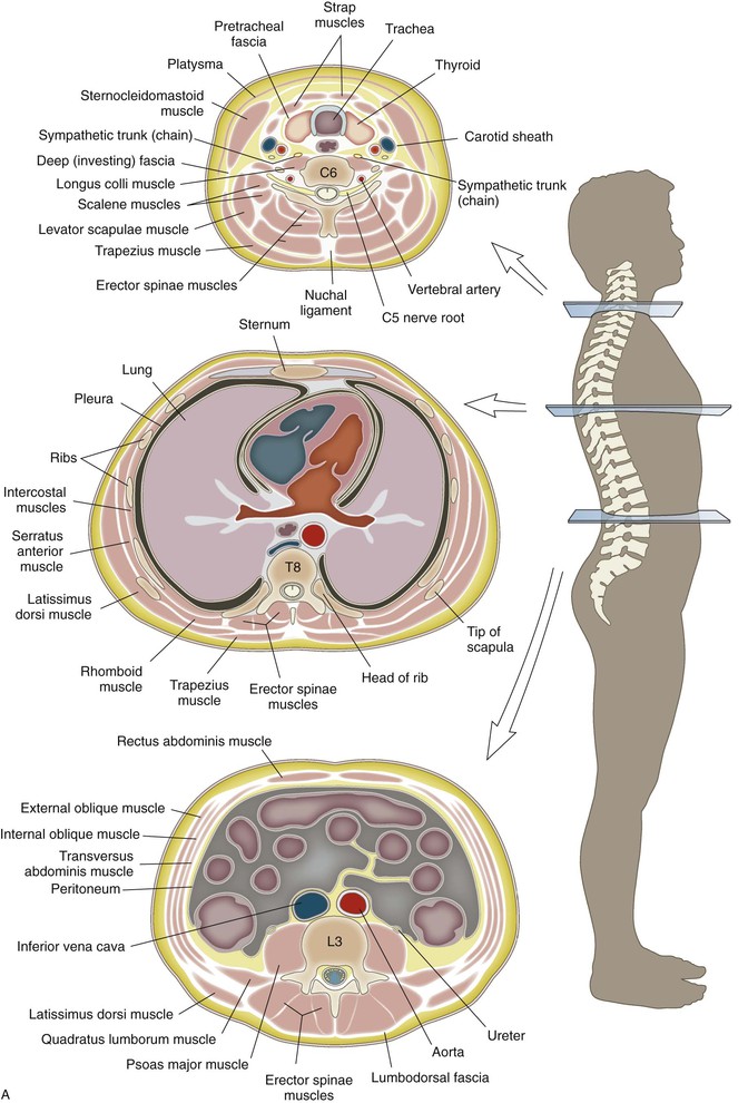
 Cross Sectional Anatomy An Overview Sciencedirect Topics
Posting Komentar
Posting Komentar