Above the knee the sciatic nerve divides into two major nerves the tibial nerve and the common peroneal nerve. These two nerves travel to the lower leg and foot supplying sensation and muscle control.
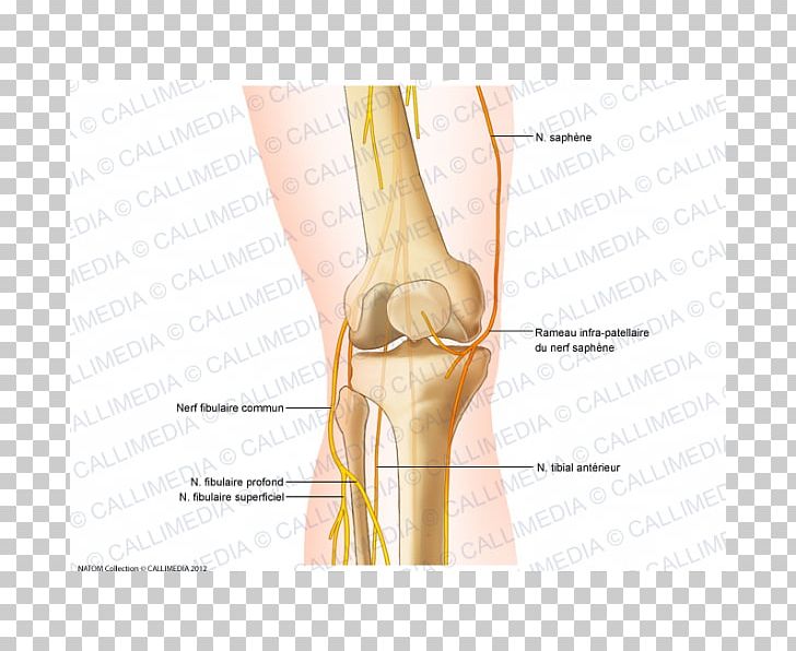 Knee Common Peroneal Nerve Anatomy Saphenous Nerve Png
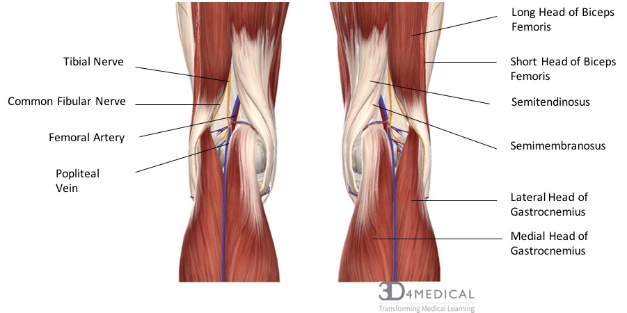
The most important nerves around the knee are the tibial nerve and the common peroneal nerve in the back of the knee.

Nerves in knees anatomy. Name location and functions of knee legaments. Each type of nerve is the relay for pain signals that originate in a different area of your knee. The tibial nerve runs downward in the midline and passes between the two heads of gastrocnemius along with the popliteal vessels.
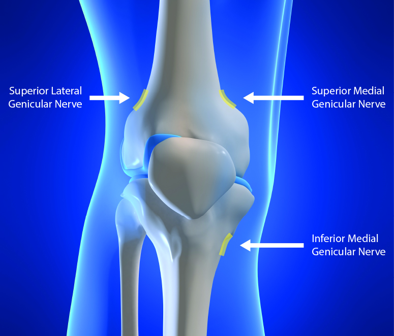
These nerves are broadly known as genicular nerves. The most important nerves around the knee are the tibial nerve and the common peroneal nerve in the back of the knee. The longest and the heaviest bone in the body.
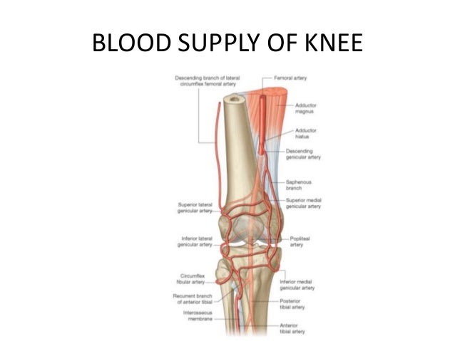
The nerve supply to the knee is derived from. Bones of the knee fig1. With the knee bent a doctor can pull anterior drawer test and push posterior drawer test the lower leg while holding the foot stable to check the stability of the acl and pcl.
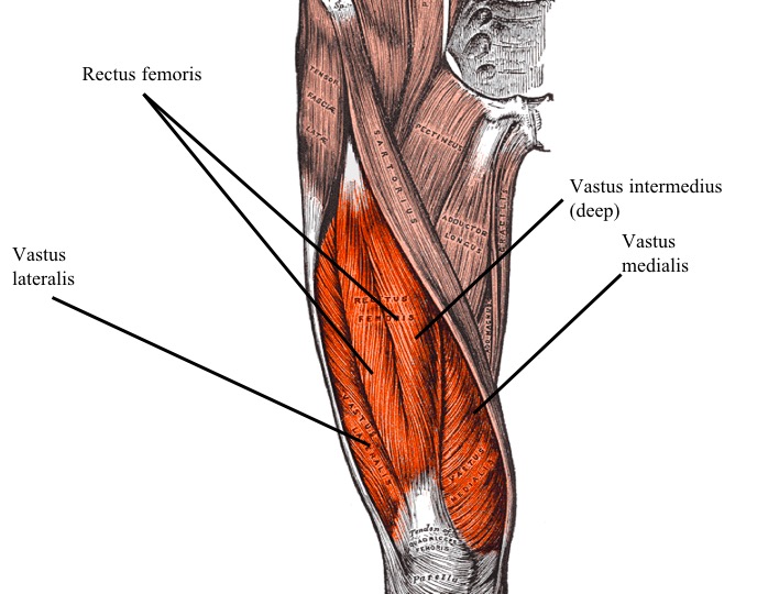
Muscles are important contributors the knee joint stability. Branches of the femoral nerve to vastus medialis and also intermedius and lateralis from the sciatic by genicular branches of the tibial and common peroneal nerves. Various nerves and blood vessels supply the muscles and bones of the knee.
The thigh bone femur the shin bone tibia knee cap patella and the fibula see image to the left. It has branches that serve the muscles of the posterior compartment before moving down toward the ankle and foot. Anatomy of the knee bones muscles arteries veins nerves bone and ligaments.
The following nerves of the popliteal fossa and leg. These two nerves travel to the lower leg and foot supplying sensation and muscle control. Sciatica can lead to nerve pain in the knee as well and compression on the sciatic nerve can occur anywhere from the lower back down to the lower leg.
The large sciatic nerve splits just above the knee to form the tibial nerve and the common peroneal nerve. This nerve branches off the sciatic nerve and runs down the midline of the popliteal fossa. There are four bones around the knee.
Identifying where the compression is taking place can help alleviate damage due to sciatica and an examination of the lower back and the legs will probably be in order. Bones of the knee. The common peroneal nerve diverges laterally running just behind the tendon of biceps femoris.
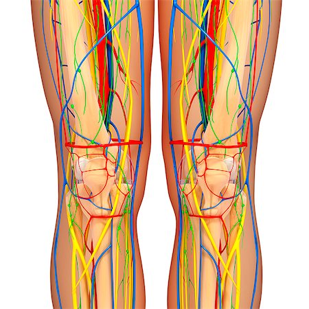
All of these different types of anatomy are crisscrossed and enervated by a network of femoral common peroneal saphenous tibial and obturator nerves.
 Knee Joint Anatomy
 Genicular Nerve Block Rf Neurotomy Of Knee Joint Pain
 Nerves Blood Vessels And Lymph Advanced Anatomy 2nd Ed
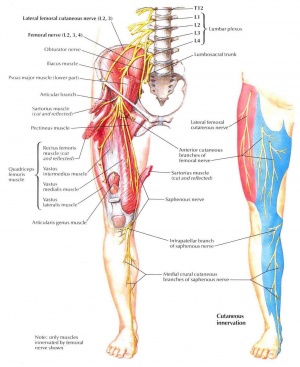 Femoral Nerve Physiopedia
 Knee Arthroscopy Setup Diagnosis Portals And Approaches
 Surface Anatomy Advanced Anatomy 2nd Ed
 Knee Joint Hanna Joints Hannakyleighmorgankaci
 Knee Anatomy Nerves Stock Photos Page 1 Masterfile
 Genicular Neurotomy Nerve Ablation Ainsworth Institute

 Knee Wikipedia
Nerves Of The Leg And Foot Interactive Anatomy Guide
 Untitled Document
 Anatomy Of The Knee Bones Muscles Arteries Veins Nerves
 The Knee Patella Tendinopathy
 Ultrasound Guided Obturator Nerve Block Nysora
 Functional Regional Anesthesia Anatomy Nysora

 Thumb Knee Nerve Human Leg Human Anatomy Knee Pain
 Patella Approach Mid Axial Longitudinal Approach Ao
 Anatomy Of The Saphenous Nerve Download Scientific Diagram
 01 Knee Anatomy

 Nerves Anatomy Exhibits
 Baker S Cysts An Early Indication Of Pathology
 Anterior Knee Pain Kennedy Brothers Physical Therapy


Posting Komentar
Posting Komentar