Osteology elbow is a hinged joint. The shaft is comprised of compact bone containing a medullary canal traversing its length.
 Distal End Of Humerus Anatomy Diagram Quizlet
When struck it can cause a distinct tingling sensation and sometimes a significant amount of pain.

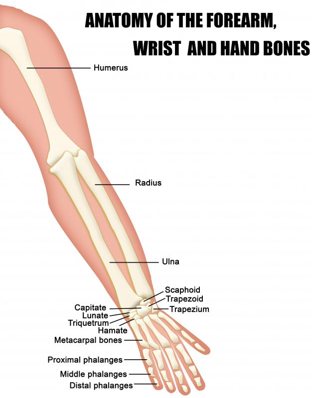
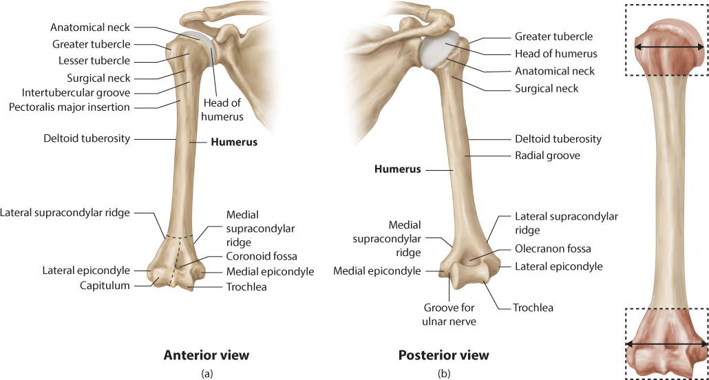
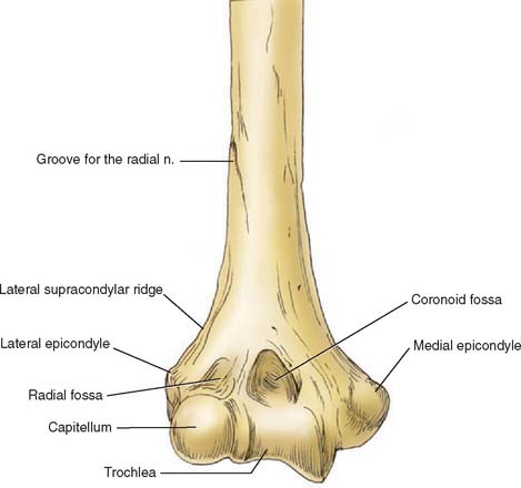
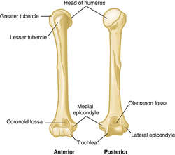
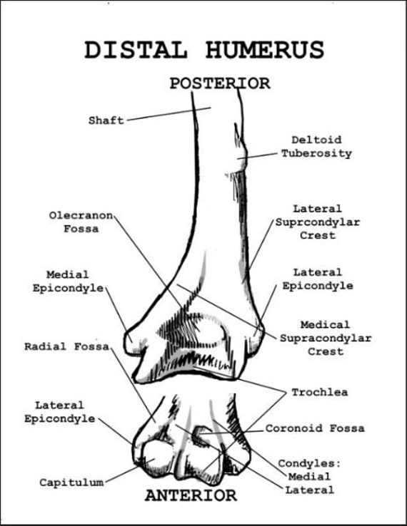
Distal humerus anatomy. The top of the arm bone is called the proximal humerus and the bottom of the bone is called the distal humerus. It extends from your shoulder to your elbow where it joins with the ulna and radius bones of your forearm. The base of the triangle comprises the transverse condylar mass.
The proximal and distal ends of the humerus are cancellous bone with a superficial layer of compact bone. Humerus fractures are generally divided into three types of injuries based on the location of the fracture. A humerus fracture is an injury to the bone of the upper arm that connects the shoulder to the elbow.
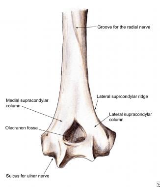
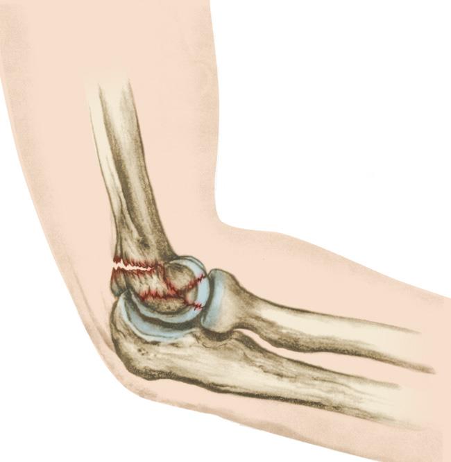
At the midshaft of the humerus the radial nerve travels from the posterior to the anterior aspect of the bone in the spiral groove. Allows for flexion and extension. A distal humerus fracture is a break in the lower end of the upper arm bone humerus one of the three bones that come together to form the elbow joint.
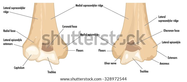
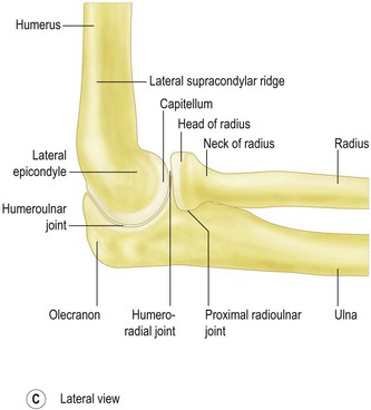
The difficulty in treating complex distal humerus fractures lies in. This mass is composed of the lateral epicondyle the capitellum the trochlea and the medial epicondyle. A humerus fracture refers to any break in this bone.
A fracture of the humerus in this region can result in radial nerve injury. The incidence of fractures of the elbow joint is small compared with that. A fracture in this area can be very painful and make elbow motion difficult or impossible.
The bones have moved out of place displaced fracture pieces of bone have punctured the skin open fracture because of the increased risk of infection open fractures are scheduled for surgery as soon as possible usually within hours. In the child the distal humerus is an osteochondral block of roughly triangular shape attached to the distal humeral diaphysis. The following nerves are located on the following aspects of the humerus.
Surgery is usually required for distal humerus fractures in which. The pain from a humerus fracture often extends to either your shoulder or elbow depending on where the break is and recovery may last several weeks. Muscles common flexors originate from medial epicondyle pronator teres.
Distal humerus fractures practice essentials. Distal humerus fractures are traumatic injuries to the distal part. Trochlea articulates with sigmoid notch.
The ulnar nerve lies at the distal end of the humerus near the elbow. Capitellum articulates with proximal radius allows for forearm rotation.
 What Is The Distal Humerus With Pictures
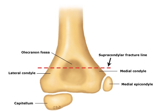
 Supracondylar Humerus Fractures Practice Essentials
![]() Humerus Anatomy And Clinical Notes Kenhub
Humerus Anatomy And Clinical Notes Kenhub
Distal Humerus Fractures Trauma Orthobullets
 Humeral Diaphyseal Fractures Musculoskeletal Key
 Elbow Arm Anatomy
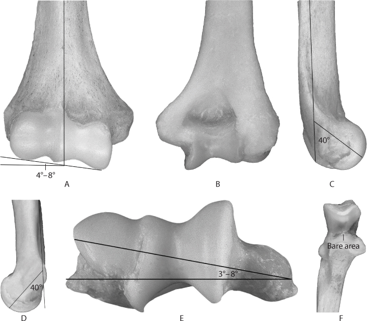
 Distal Humerus Showing Major Anatomical Features Royalty
Plos One The Course Of The Radial Nerve In The Distal
 The Distal Humerus Showing The Major Anatomical Features And
 Anatomy Of The Elbow Joint Clinical Gate
 Humerus Definition Of Humerus By Medical Dictionary
 Distal Humerus Fractures Of The Elbow Orthoinfo Aaos
 Amazon Com Vision Scientific Vaj234 Classic Functional
 Crackcast E052 Orthopedics Humerus And Elbow Canadiem
 Fractures Of The Distal Humerus Ppt Download
 Distal Humerus Approach Distal Humeral Anatomy E 2 1
 Elbow Anatomy Musculoskeletal Key
 Startradiology
 Distal Humerus O Nil 3 5 Series Intrauma S P A
 Labeled Distal Human Humerus Bone Anatomy Wall Art Jpeg Download
 The Management Options For Adult Distal Humeral Fractures
 The Elbow
 Pediatric Supracondylar Fractures Core Em
 Distal Humeral Fractures Musculoskeletal Key


Posting Komentar
Posting Komentar