Normal mri anatomy of the knee 639 the fcl biceps femoris bursa is found lateral to the distal fcl and insinuates anterior and antero medial in relation to this ligament. Mri knee joint anatomy.
Fractures Of The Proximal Tibia Shinbone Orthoinfo Aaos
Colorado knee specialist dr.

Mri knee anatomy. Anatomy of the knee can be complicated and hard to understand. Proximal attachment of posterior cruciate ligament j. Medial patellar retinaculum b.
Anatomy of the knee mri atlas of the human body using cross sectional imaging. Use the mouse to scroll. 3mm 3dft fourier transformation sub centimeter 5.
Lateral tibial plateau cartilage kmedial collateral ligament partially torn in this patient l. Three conventional mri planes that are utilized to evaluate the knee include sagittal oblique coronal and transaxial planes. Patient positioning supine with the leg in full extension.
Robert laprade discusses how to read an mri of a normal knee. The knee is placed in 10 to 15 of external rotation esp for sagittal image slicethickness 3 4 mm sections are used for axial coronal and sagittal images of the knee. Anterior root attachment of medial meniscus g.
Click on a link to get t1 coronal view t2 fatsat axial view t2 fatsat coronal view t2 fatsat sagittal view. Hoffas fat pad f. This mri knee sagittal cross sectional anatomy tool is absolutely free to use.
Use the mouse scroll wheel to move the images up and down alternatively use the tiny arrows on both side of the image to move the images. Each anatomical structure is labelled interactively. Using high energy magnetic waves an mri scanner creates highly detailed images of the knee and leg.
This atlas of cross sectional anatomy of the knee is based on imagery by magnetic resonance mri. Trochlear groove cartilage e. Through the use of magnetic resonance imaging clinicians can diagnose ligament and meniscal injuries along with identifying cartilage defects bone fractures and bruises.
While a detailed explanation of mri protocols and mr physics is beyond the scope of this text fast spin echo fse mri is most commonly utilized for mri of the knee. Atlas of knee mri anatomy. This webpage presents the anatomical structures found on knee mri.
Magnetic resonance imaging mri scan. This tool is at the same time useful for the training and teaching of the anatomy. Iliotibial band hanterior horn of lateral meniscus i.
An mri scan is the most often used method of detecting.
 Department Of Anatomy Med Univ Of Warsaw Poland Knee

 James Y Song Msiv Gillia
 Mri Knee Anatomy
 How To Read Knee Mri Of Normal Knee Anatomy Of The Knee Colorado Knee Specialist
 Mri Knee Joint Anatomy
 Knee Mri Approach To Msk Mri Series
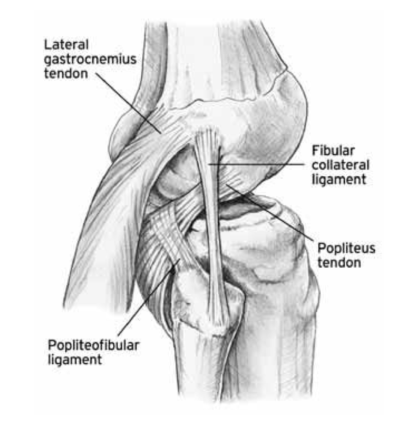 The Knee Resource Posterolateral Corner Injury
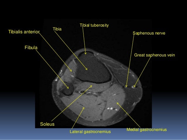
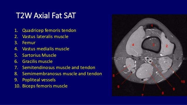
 Mri Anatomy Of Knee Dr Muhammad Bin Zulfiqar
 Knee Wikipedia
 Knee Mri Sequences
 Knee Anatomy Mri Knee Coronal Anatomy Free Cross
 Normal Acl Pcl Left Knee Anatomy Ruptured Acl Mri Of
 Mri Knee Joint Anatomy
 Mri Anatomy Of The Knee And Shoulder Lieberman S
 Imaging Of Tumors And Tumor Like Lesions Of The Knee
 Module 2 Lower Extremity Orthopedic Imaging
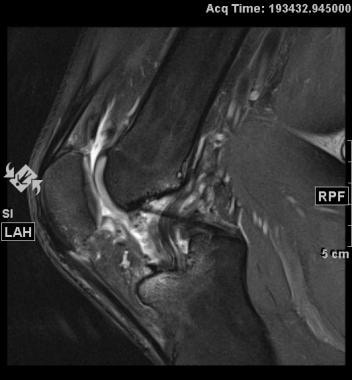
 Essr 2015 P 0055 Popliteal Fossa Masses A Pictorial

 Mri Anatomy Of Knee Dr Muhammad Bin Zulfiqar
 Mri Knee Anatomy Knee Sagittal Anatomy Free Cross

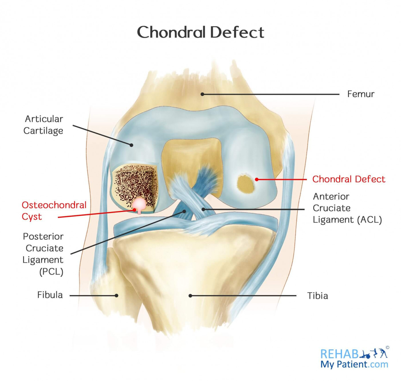 Chondral Defect Rehab My Patient
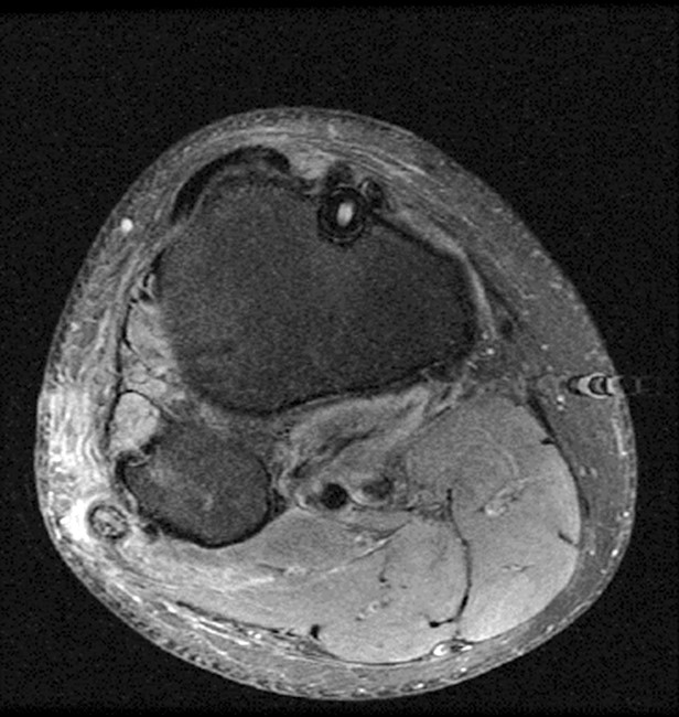
 Mri Knee Sagittal Anatomy Quiz Radiology Case
 The Radiology Assistant Knee Non Meniscal Pathology
 Figure 14 From Normal Mr Imaging Anatomy Of The Knee
 A Guide To Your Knees Well Guides The New York Times
Stanford Msk Mri Atlas C 2019

 Mri Of The Whole Body An Illustrated Guide For Common
![]() Medical Imaging And Radiological Anatomy X Ray Ct Mri
Medical Imaging And Radiological Anatomy X Ray Ct Mri
 Mri For Anterior Cruciate Ligament Injury Overview Anatomy


Posting Komentar
Posting Komentar