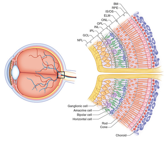
Simple anatomy of the retina by helga kolb. The optic nerve contains the ganglion cell axons running to the brain and additionally incoming blood vessels that open into the retina to vascularize the retinal layers and neurons fig.
 High Yield Topic Clinical Anatomy Of Retina
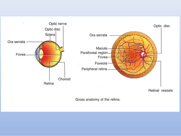
The retina is approximately 05 mm thick and lines the back of the eye.

Anatomy of retina. The neural retina consists of several layers of neurons interconnected by synapses and is supported b. This page describes normal retinal anatomy. The retina is the innermost light sensitive layer of tissue of the eye of most vertebrates and some molluscs.
This fundus photograph shows the normal appearance of the retina. The red curving structures are blood vessels which enter the retina through the nerve. Anatomy of retina by drashok kumar valuroutu 2.
From the center of the optic nerve radiate the major blood vessels of the retina. This is a small tube that runs from the eye to the nasal cavity. In the diagram above anatomy of the eye the artery is shown in red while the vein is shown in blue.
The retina is a thin layer of tissue that lines the back of the eye on the inside. The retina is a thin semitransparent multilayered sheet of neural tissue that lines the inner aspect of the posterior two thirds of the wall of the globe. The retina processes light through a layer.
Refer to this page for comparison with the retinal disease pages. It is located near the optic nerve. The whitish circle is the nerve that connects the retina to the brain.
The purpose of the retina is to receive light that the lens has focused convert the light into neural signals and send these signals on to the brain for visual recognition. In the center of the retina is the optic nerve a circular to oval white area measuring about 2 x 15 mm across. The central artery and vein runs through the center of the optic nerve.
The central artery supplies the retina while the central vein drains the retina. The optics of the eye create a focused two dimensional image of the visual world on the retina which translates that image into electrical neural impulses to the brain to create visual perception the retina serving a function analogous to that of the film or image sensor in a camera.
 Eye Conditions Florida Eye Clinic
 Anatomy Of Retina
 Retina Vitreous Anatomy
 Anatomy Of Retina
 Pdf Retinal Anatomy And Pathology
 Blind Spot Anatomy Britannica
 Retinal Anatomy
 Anatomy Of The Eye And Arrangement Of Cells In The Retina
 Anatomy Crystal Clear Eye Surgeons
 Anatomy Of The Eye Kellogg Eye Center Michigan Medicine

 Anatomy Of The Eye Moorfields Eye Hospital

 Anatomy Of The Eye Retina Ophthalmologist Gettysburg Pa
The Anatomy Of Retina From Outer Layer Up To Inner Layer
 Anatomy Of Retina
 Anatomy Of Retina
 Anatomy Of Retina
 Retina Anatomy Physiology
 Glossary Anatomy Of The Eye

:max_bytes(150000):strip_icc()/GettyImages-308783-003-56acdcd85f9b58b7d00ac8e8.jpg) The Anatomy Of The Retina
The Anatomy Of The Retina
 Retina Anatomy And Physiology
 Midterm Spring Anatomy Physiology Lab With Nelson At
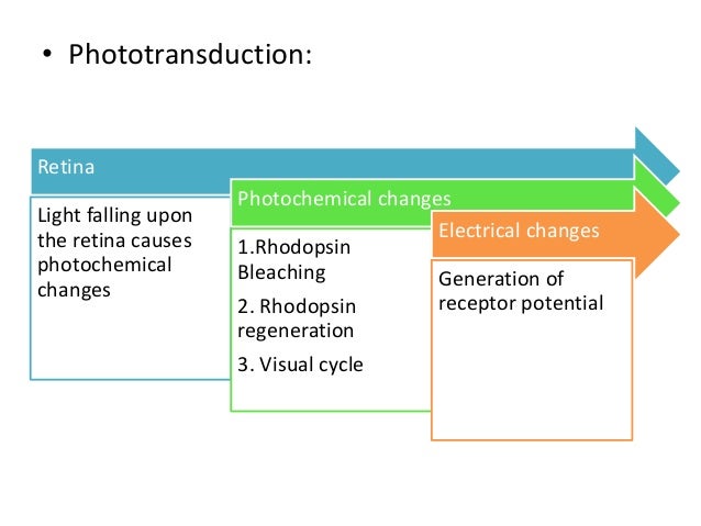
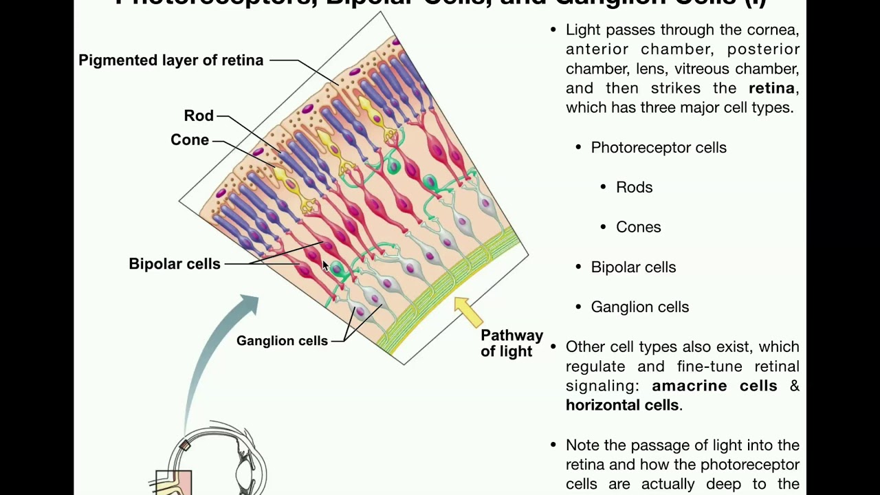 Anatomy Vision Part 1 Retina Photoreceptors Bipolar Cells Ganglion Cells


Posting Komentar
Posting Komentar