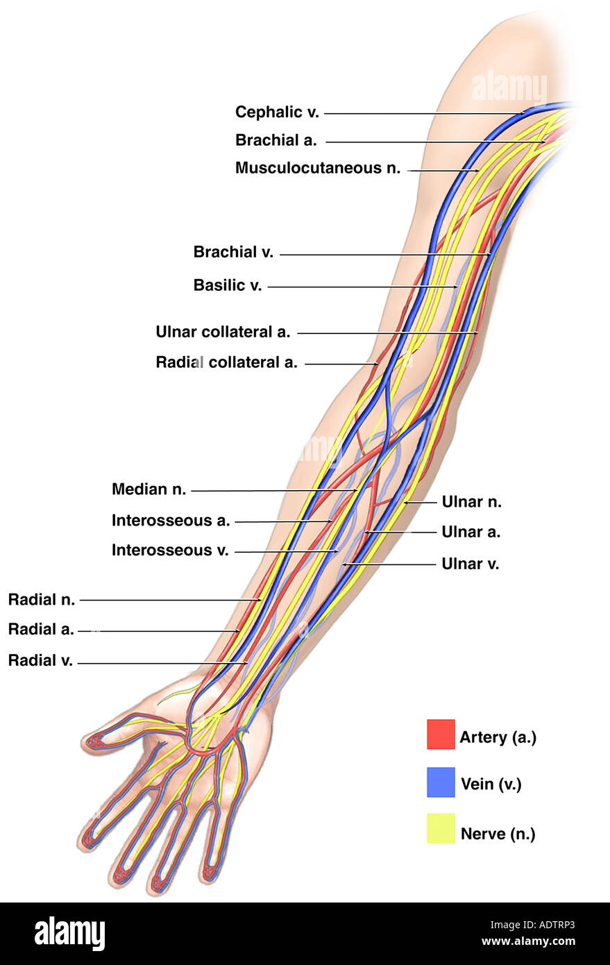
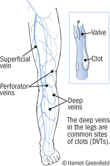
Deep veins and superficial veins. The pulsations of the brachial artery assist the venous return.
 Vein Elbow Forearm Anatomy Artery Png Clipart Abdomen
The patient will be comortable seated with a pillow under their arm for support and this allows for easy scanning.
Arm vein anatomy. Arm like in the forearm the arm is drained by the brachial veins deep veins that accompany the brachial artery and all its branches. Veins that are structured in this way are known as vena comitantes. An axial view of the upper arm vein anatomy.
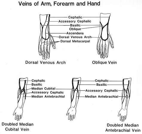
The veins of the arm carry blood from the extremities of the limb as well as drain the arm itself. The veins of the arm may be divided into two groups. Transverse view of the brachial and basilic veins and nerves of the upper arm.
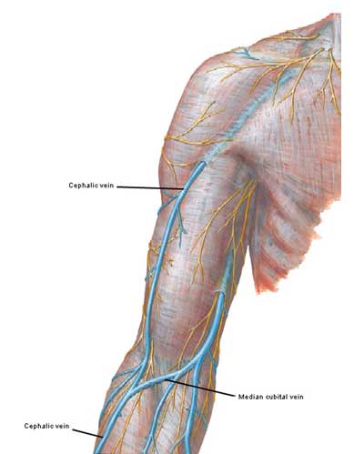
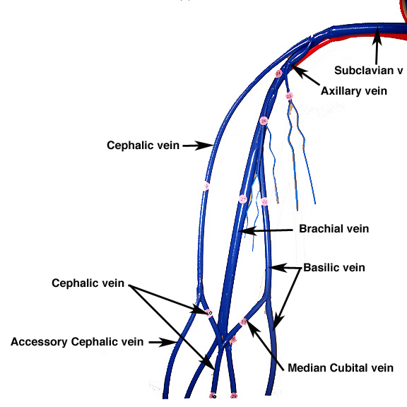
Specifically the main veins of this area are the deep brachial veins deep veins that accompany the brachial artery and the basilic and cephalic veins. The brachial veins are the largest in size and are situated either side of the brachial artery. The two main veins are the basilic and the cephalic veins.
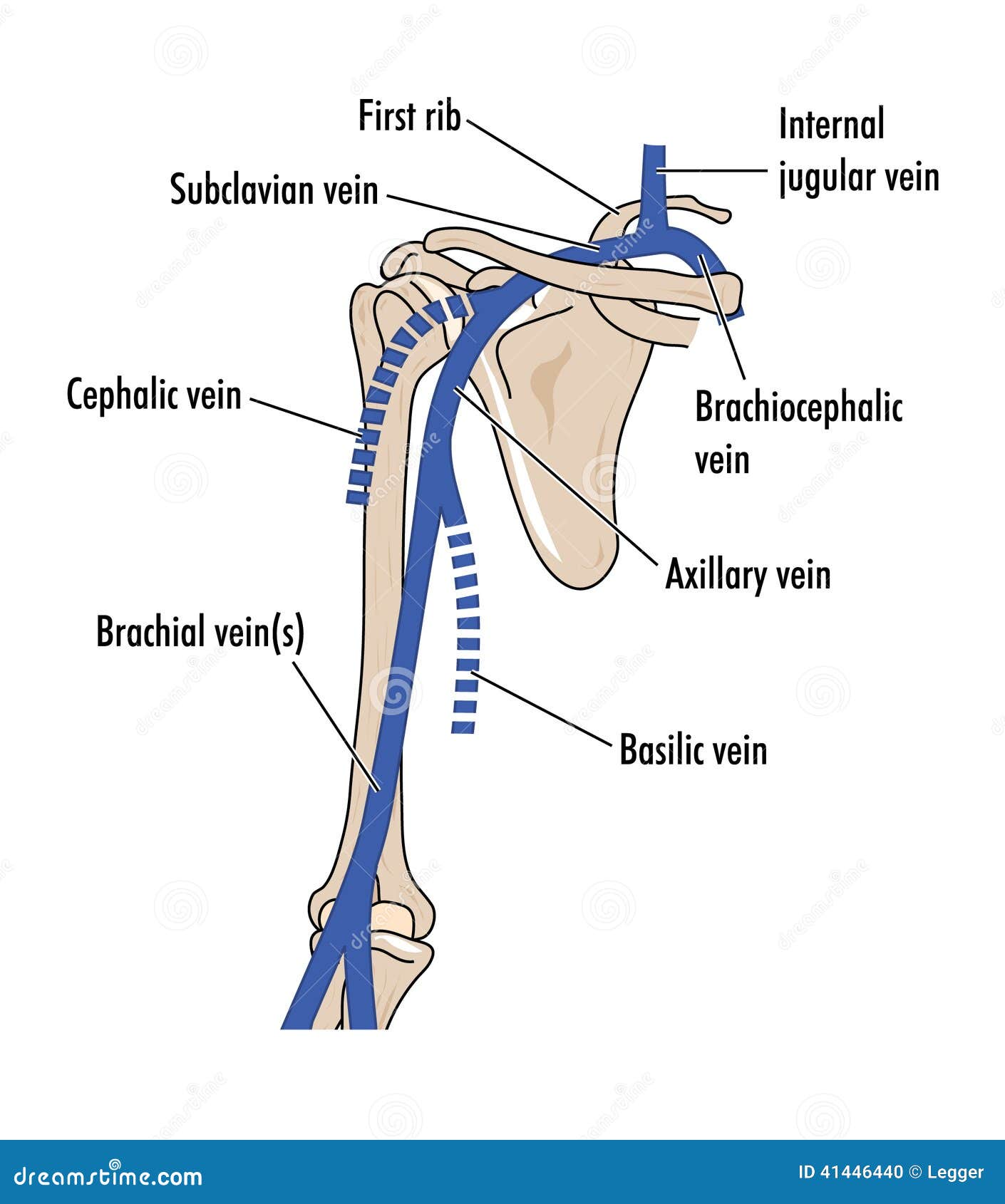
There is a connecting vein between the two the median cubital vein which passes through the cubital fossa and is clinically important for venepuncture withdrawing blood. Veins of the upper limb the primary venous return from the arm is through the axillary vein which continues centrally as the subclavian and brachiocephalic innominate veins before emptying into the superior vena cava. The venous drainage of the arm is a continuation of the venous system of the forearm.
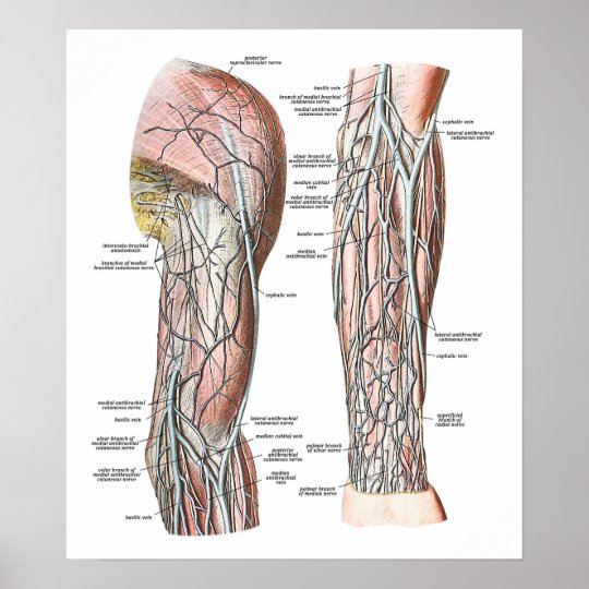
Opposite the cephalic vein the basilic vein travels through the arm near the triceps muscle on the underside of the arm. All these veins drain finally to the subclavian veins. Damage to these major veins and arteries especially trauma.
The veins will be easier to assess with the patient erect to allow better venous distension. Superficial veins of the upper limb in the cadaver. Perforating veins run between the deep and superficial veins of the upper limb connecting the two systems.
 Frank Netter Page 8 Outlander Anatomy
 Veins Of The Arm Anatomy Study Buddy

 Lower Extremity Venous Anatomy Dallas Tx Venous System
![]() Cephalic Vein Anatomy And Clinical Points Kenhub
Cephalic Vein Anatomy And Clinical Points Kenhub
 Cephalic Vein Wikipedia
 Anatomy Veins Of The Hand And Forearm Critical Care
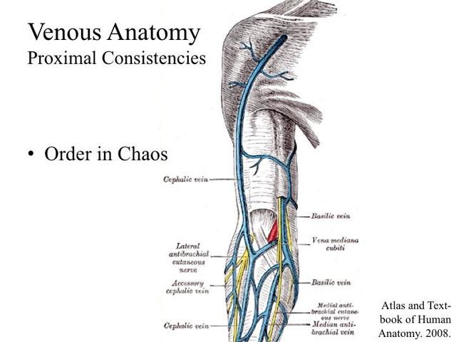

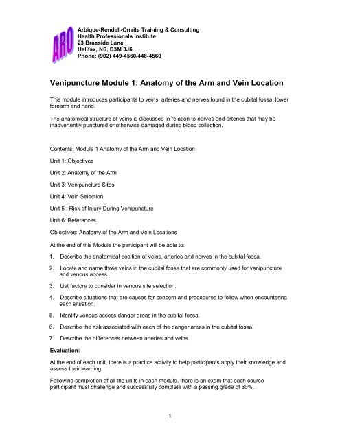 Venipuncture Module 1 Anatomy Of The Arm And Vein Location
Veins Of The Upper Arm Stock Vector Illustration Of Humerus
 Veins Of The Arm Poster
 Instant Anatomy Diagram
 Anatomy Of The Nerves Arteries And Veins Of The Arm Upper
 Upper Arm Vein Anatomy Arm Veins Arm Anatomy Upper Limb
![]() Veins Of The Upper Limb Anatomy Kenhub
Veins Of The Upper Limb Anatomy Kenhub
 Vein Wikipedia
 Forearm Artery And Venous System
 Fig Forearm And Hand Arterial And Venous Anatomy With The
 Anatomy Of The Nerves Arteries And Veins Of The Arm Upper
 Anatomy Atlases Illustrated Encyclopedia Of Human Anatomic
 Cardiovascular System Of The Arm And Hand

 Arm Vein Anatomy Diagram Quizlet
 Deep Vein Thrombosis Blood Clots In Your Veins Harvard Health
 Arm Dvt Normal Ultrasoundpaedia


Posting Komentar
Posting Komentar