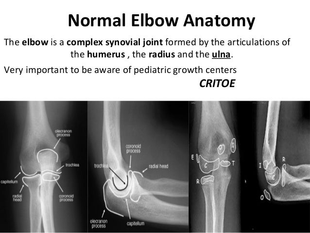
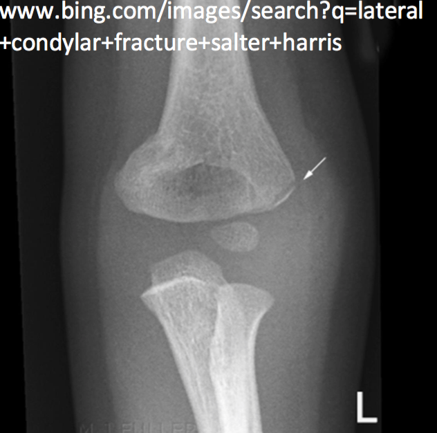
Gross anatomy articulations the elbow joint is made up of three articulations 23. This anatomy is increasingly important in evaluating abnormalities such as osteonecrosis of the capitellum panners disease osteochondral defects and medial apophysitis little league elbow for example.
 Film Xray Elbow Radiograph Show Normal Stock Photo Edit Now
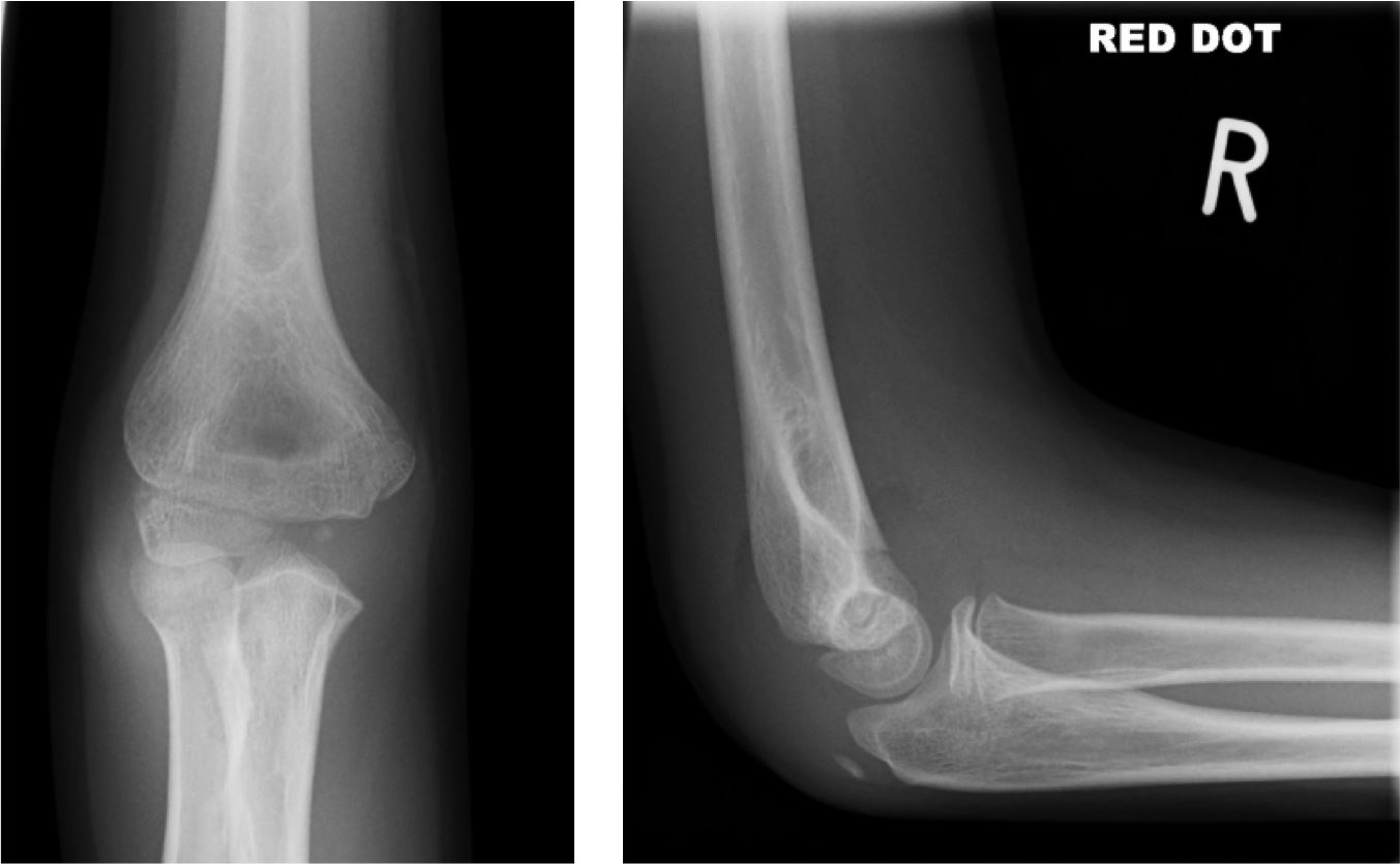
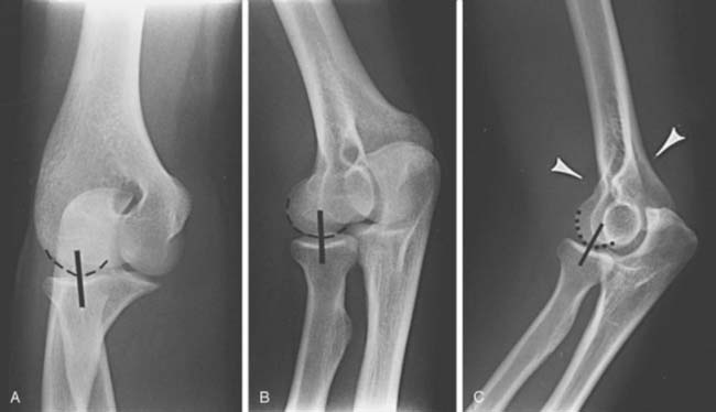
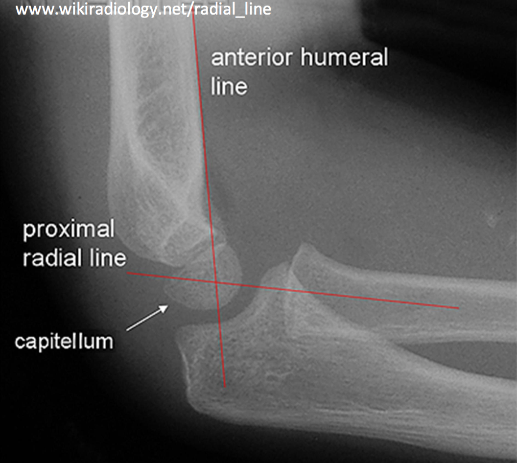
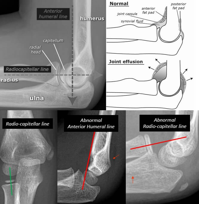
Alignment fat pads bone cortex alignment check the anterior humeral line.

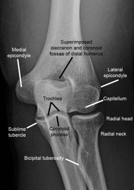
Elbow xray anatomy. Systematic review whenever you look at an adult elbow x ray review. When you study the anatomy of the elbow it is good to use the inside out approach. Test your knowledge about elbow xray anatomy with this online quiz.
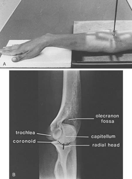
The anterior fat pad is seen in most but not all normal elbows. However as emergency physicians it is important to recognize the elbow ossification centers that develop during childhood in order to accurately interpret radiographs of the joint. Place the arm on the table with elbow straight.
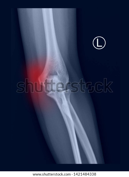
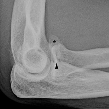
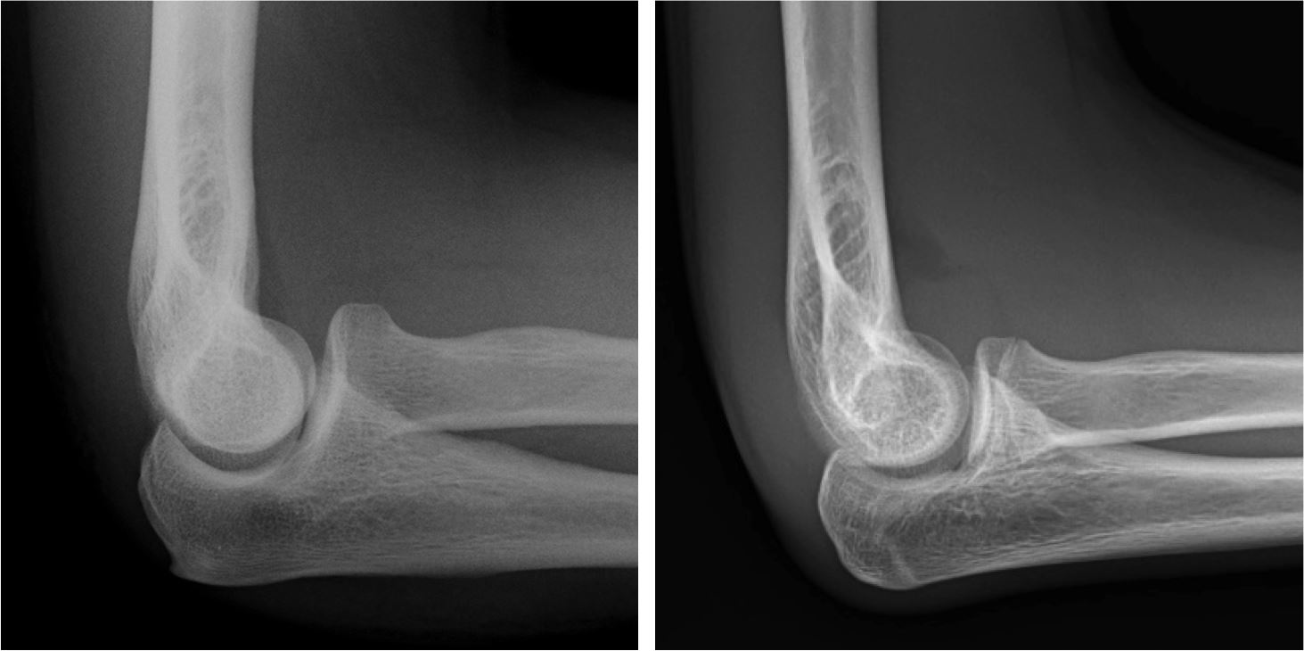
Elbow fat pads there are pads of fat close to the distal humerus anteriorly and posteriorly. Normal elbow x ray appearances on the lateral image there is often a visible triangle of low density lying anterior to the humerus. This is a normal structure.
Drawn down the anterior surface of the humerus should intersect the middle 13 of the capitellu. Capitellum of the humerus with the ra. Normal ossification centers of the elbow.
First study the bones and then continue with the ligaments and the tendons and then the surrounding structures. Stanford bone tumor bayesian network issssr msk lectures for residents ocad msk cases from around the world stanford msk mri atlas has served almost 800000 pages to users in over 100 countries. They are extrasynovial but intracapsular.
The elbow is a complex synovial joint formed by the articulations of the humerus the radius and the ulna. In order to establish treatment algorithms and evaluate outcomes common and reliable methods of measurement and assessment are necessary. A trivia quiz called elbow xray anatomy.
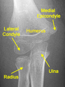
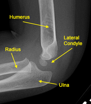
Ideally the upper arm elbow and forearm are all resting on the table. We all know the basic anatomy of the elbow with the humerus radius and ulna. Position of part place the arm on the table with the elbow straight and the palm of hand straight up.
Ideally the upper arm elbow and forearm are all resting on the table. This is the anterior fat pad which lies within the elbow joint capsule.
 The Elbow
 Startradiology

 Diagnostic Imaging Of The Elbow Clinical Gate
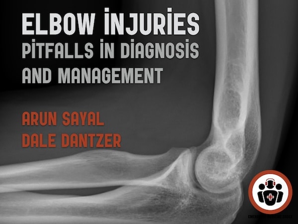 Ep 121 Elbow Injuries Ten Pitfalls In Diagnosis And
 Elbow Xray Positioning Ap Oblique Projection Lateral
 The Radiology Assistant Elbow Fractures In Children
 Diagnostic Imaging Of The Elbow Clinical Gate
 Film Critique Of The Upper Extremity Part 2 Elbow And Forearm
 Elbow Injuries Rcemlearning

 Radiology Of The Elbow Joint Dr Sumit Sharma
Elbow Olecranon Fractures Orthoinfo Aaos
 Radiological Anatomy Of The Shoulder Arm Elbow Forearm
 The Radiology Assistant Elbow Fractures In Children
 Radiologic Evaluation Of The Elbow Fundamentals Of

 Imaging Of Elbow Fractures And Dislocations In Adults
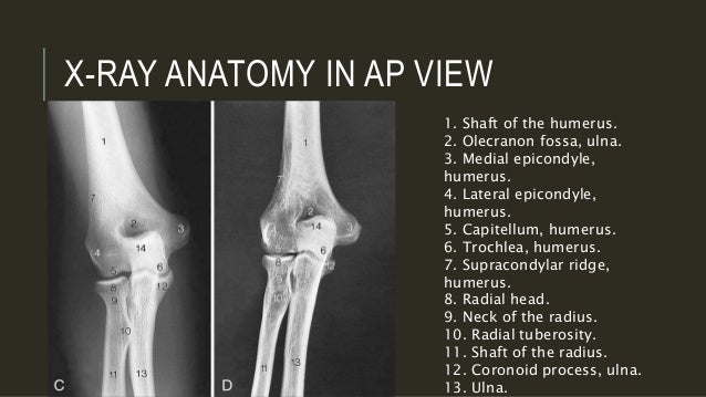
 Xray Ap Elbow Anatomy Diagram Quizlet
 Interpreting Elbow And Forearm Radiographs Taming The Sru
 Interpreting Elbow And Forearm Radiographs Taming The Sru
 Radiology In Ped Emerg Med Vol 1 Case 12
 The Radiology Assistant Elbow Mri
 Normal Radiographic Anatomy Of The Elbow Radiology Case
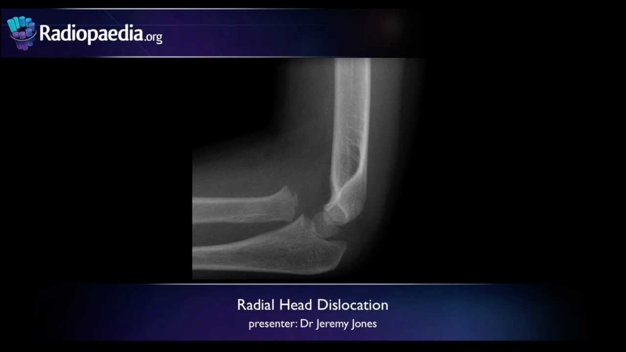 Radial Head Dislocation Radiology Video Tutorial X Ray

 Imaging Of Elbow Fractures And Dislocations In Adults
 Mnemonic Approach To Elbow Xray Fool Epomedicine
 The Elbow
 Interpreting Elbow And Forearm Radiographs Taming The Sru
 X Ray Of Elbow Joint
 Radiological Anatomy Of The Shoulder Arm Elbow Forearm


Posting Komentar
Posting Komentar