Dog front hind leg injury. Two thirds of a dogs body weight is carried on their front legs.
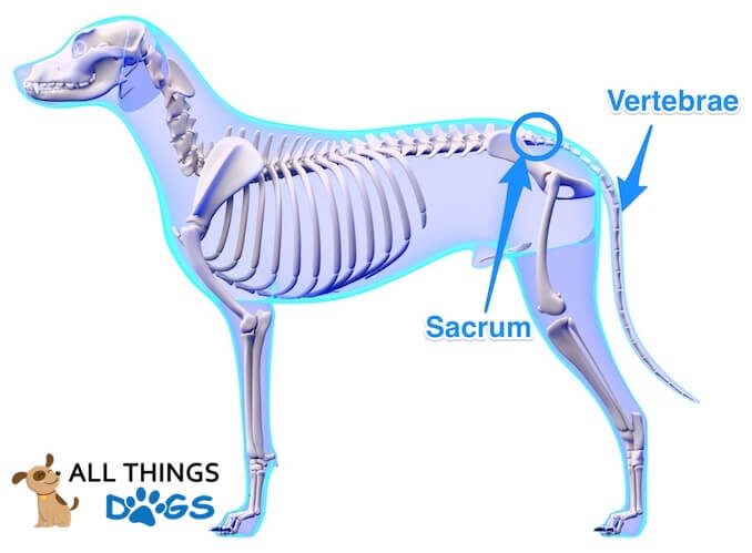
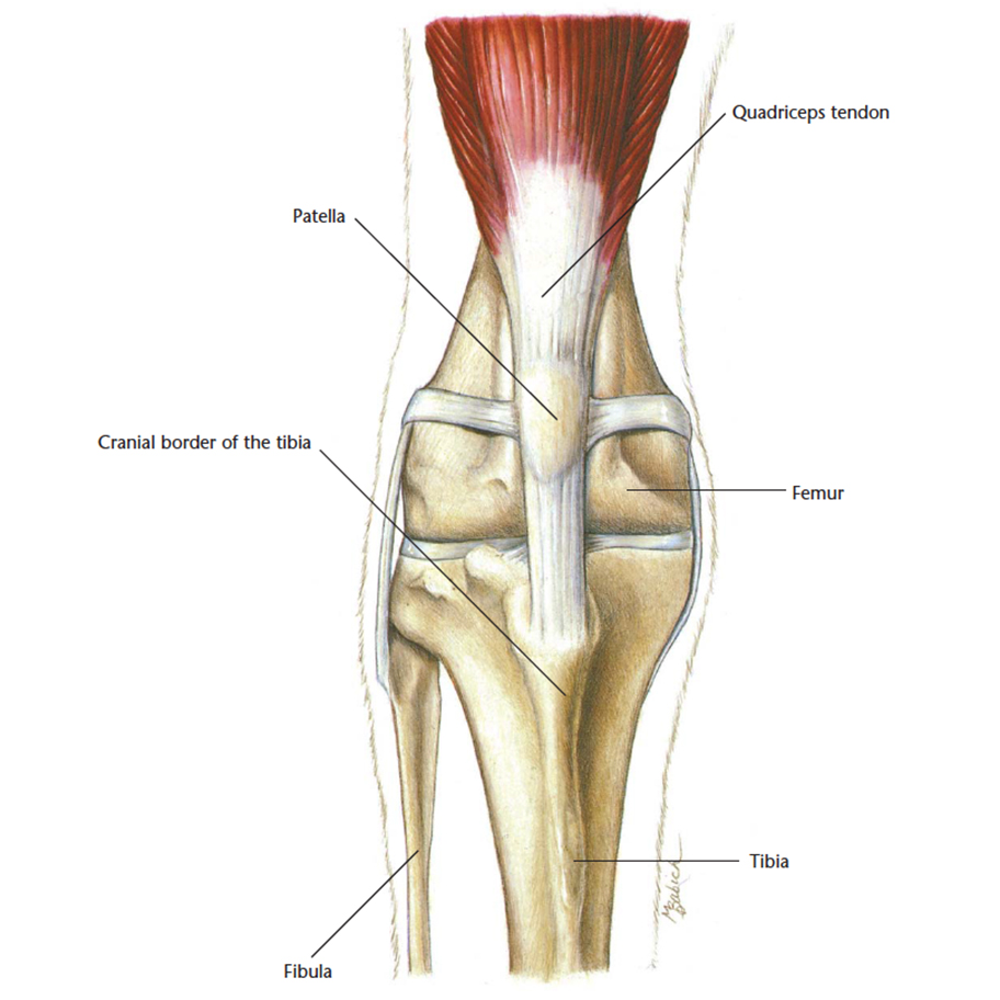
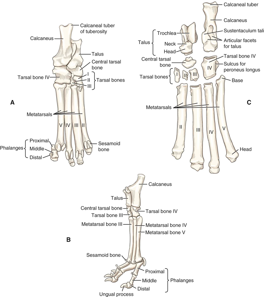 Canine Anatomy Veterian Key
The implications of front leg injuries are sometimes different from dog hind leg injury.
Dog hind leg anatomy. Hind leg anatomy images 28 images parts of human hind limb anatomy human leg images leg anatomy www pixshark images image gallery leg bones cat hind leg anatomy image collections human. Dog hind leg medial and caudal dog head legs and paws anatomy by herman dittrich vintage dog feet anatomy illustration book page by niminsshop. Only one third is carried on their hind legs.
However the muscles on their hind legs are larger and therefore stronger. The first warning sign of strains or sprains may be that your dog starts to limp or is suddenly lame meaning he cant use his leg. The anatomy of a dogs hind leg and foreleg differs just as a human arm and leg differ according to for dummies.
The dog has the greatest range of movement in this joint compared to other domestic species. Such injuries are usually characterized by severe swelling and loss of movement. The muscles affecting the pelvic girdle and hip can be divided into two distinct groups.
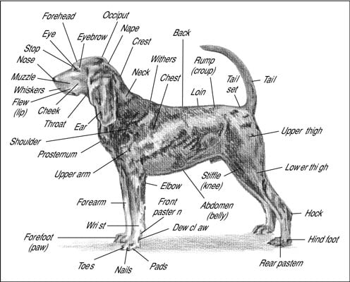
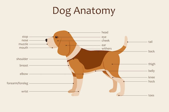
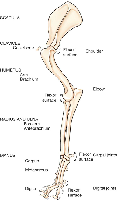
Dog leg anatomy just like humans have arms and legs dogs have forelegs and hind legs. Dogs legs are comprised of bones muscles ligaments and tendons. If this lasts more than a day or so or if it happens again and.
Causes of hind leg weakness in dogs most of the different causes are related to the dogs spinal column spinal cord or the nerves that supply the back legs. Directly below the shoulder of the foreleg is the humerus bone which ends at the elbow the first joint located just below the chest on the back of the foreleg. Injury to the spinal cord or nerves supplying the hind legs.
The girdle musculature and the rump muscles. Often called the carpals and pasterns dogs have them in both forelegs and hind legs equivalent to human bones in hands and feet excluding fingers and toes joint anatomy in dogs a joint is formed when two bones are brought together and held in place by supporting tissue. These further extend to the heel bone known as tarsus the paw bone known as metatarsus and the toe bone phalange.
Dewclaws are vestiges of thumbs. Dogs have a foot or paw at the end of each leg called the forefoot or hind foot depending on whether its front or back. They can be divided into broad categories.
The paw comes with nails sometimes called claws paw pads and usually dewclaws. If the dog has sustained muscle injuries like a sprain or has strained a muscle there may be difficulty in moving. The rear legs of the dog begin with the femur bone which extends to a pair of bones known as the tibia and the fibula.
A dogs toes are equivalent to your fingers and toes although you can wiggle yours more easily. It has the ability to flex extend rotate adduct and abduct its whole limb because of this.
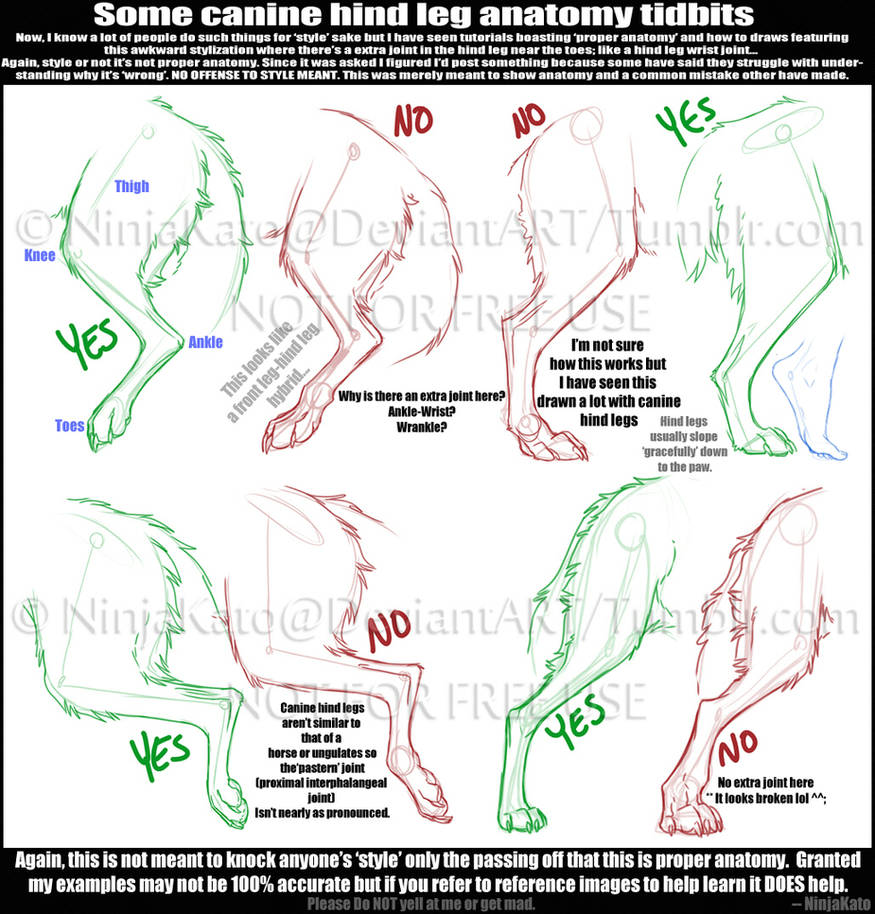 Canine Hind Leg Tidbits By Sterlingkato On Deviantart
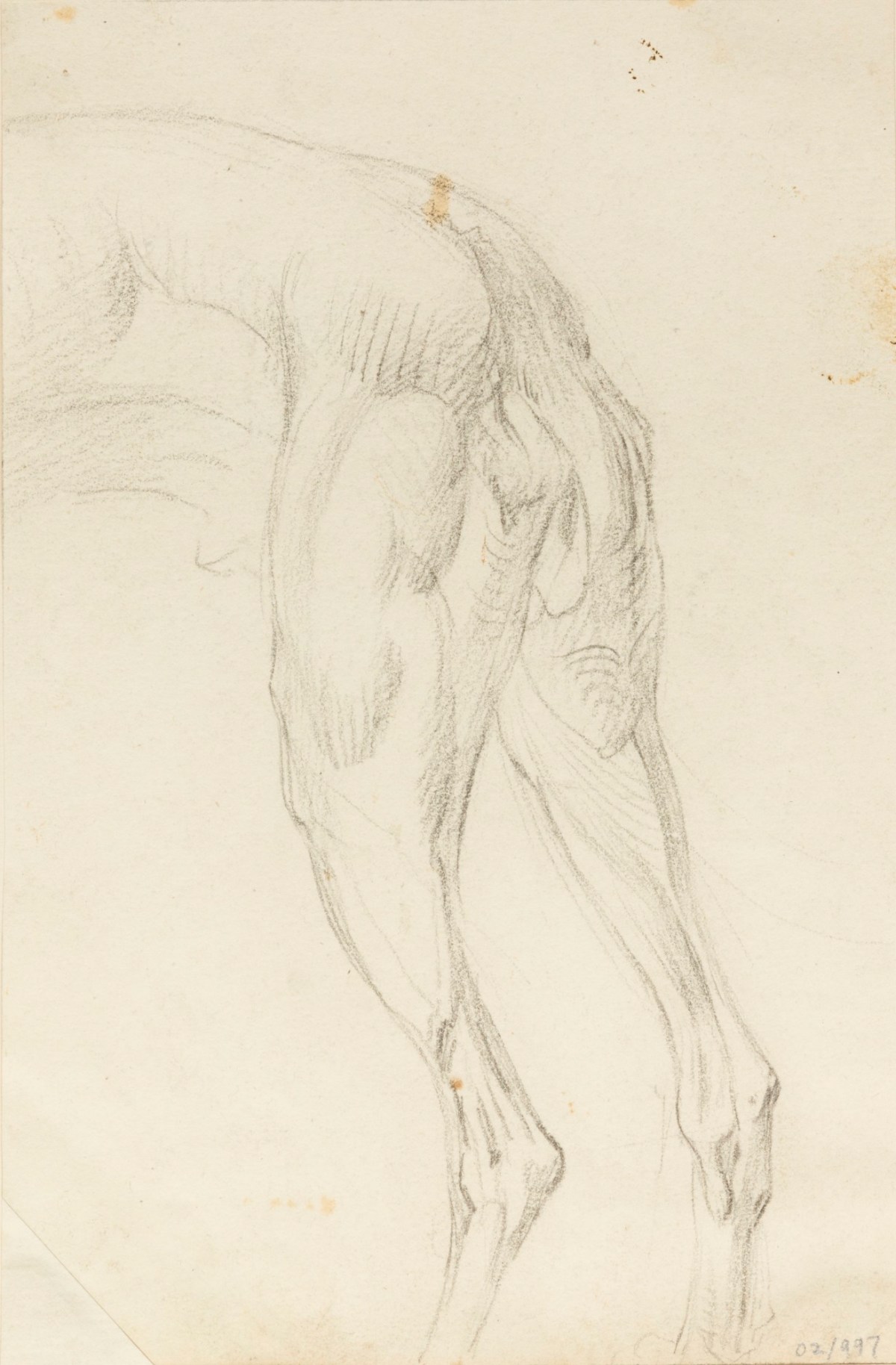 Anatomical Study Of The Muscles Of A Dog S Hind Legs Works
 Hip Dysplasia In Dogs Vca Animal Hospital
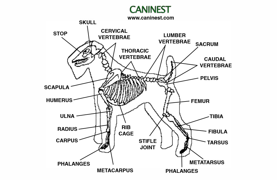
 Canine Anatomy Veterian Key
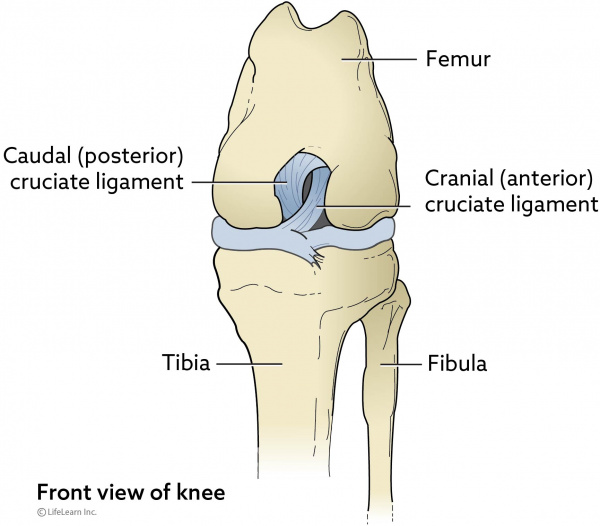
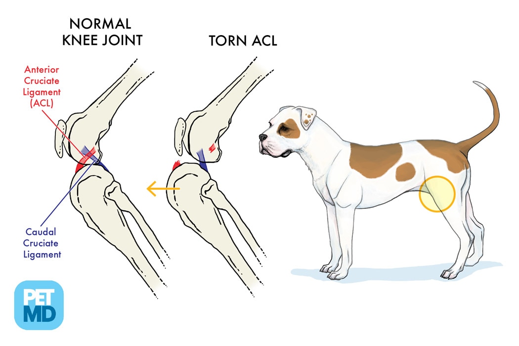 Cranial Cruciate Ligament Medical Diagram
 A Visual Guide To Dog Anatomy Muscle Organ Skeletal
 Cruciate Ligament Rupture In Dogs Vca Animal Hospital
 Dog Sprained Leg Symptoms Treatment Canna Pet
 A Visual Guide To Dog Anatomy Muscle Organ Skeletal
Dog Anatomy From Head To Tail Dummies
 Function Of The Extrinsic Hindlimb Muscles In Trotting Dogs

 The Anatomy Of Your Dog Or Cat Bothell Pet Hospital Case
 Parts Of The Nervous System In Dogs Dog Owners Merck
 Dog Anatomy Wikipedia
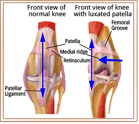 Patellar Luxation Metropolitan Veterinary Associates
 Hip Dysplasia In Dogs Treatment Diagnosis Care
 Why Your Dog Shouldn T Be Walking On Hind Legs Ultimate
 Dog Anatomy Mobility Health

 Canine Anatomy Veterian Key
 Free Art Print Of Dog Hind Legs Anatomy With Circulatory System
 Canine Hindlimb Anatomy Physiology Wikivet English
 2019 Ultimate Veterinary Guide To Dog Anatomy With Images
 Dog Anatomy Wikipedia
 Greyhound Left Front Leg And Inner Side Hind Leg Plate Dog
 Hind Leg Locking Mechanisms Horse Anatomy Horses Dog Anatomy
 Canine Anatomy Veterian Key


Posting Komentar
Posting Komentar