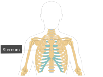
This is a division of the sternum. The thoracic cage rib cage forms the thorax chest portion of the body.
 Thorax Basicmedical Key
The thoracic cage rib cage forms the thorax chest portion of the body.
Thoracic cage anatomy. The thoracic cage takes the form of a domed bird cage with the horizontal bars formed by ribs and costal cartilages. Body of sternum what is being highlighted. Manubrium what is this.
Osteology of the thoracic cage. The thoracic cage protects the heart and lungs. Body of sternum what is this.
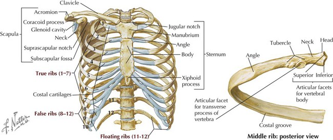
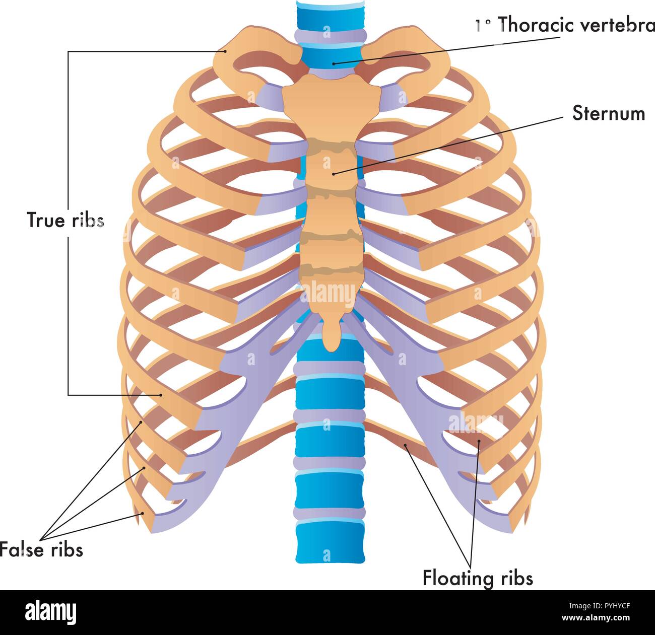
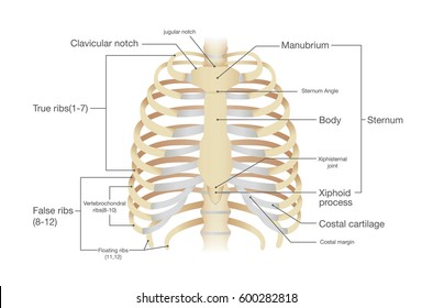
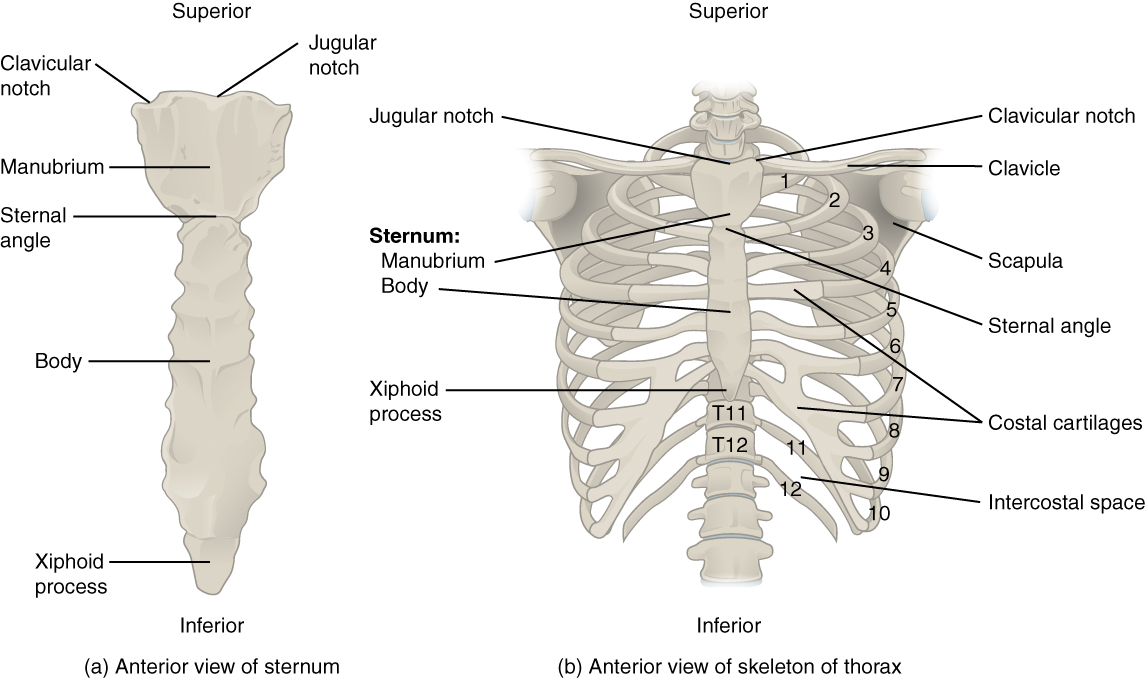
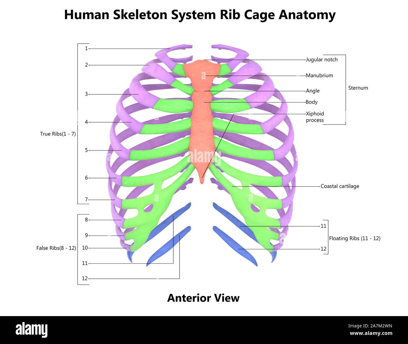
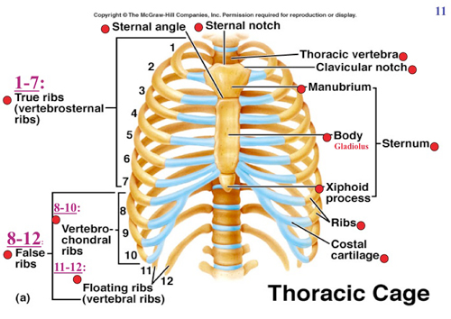
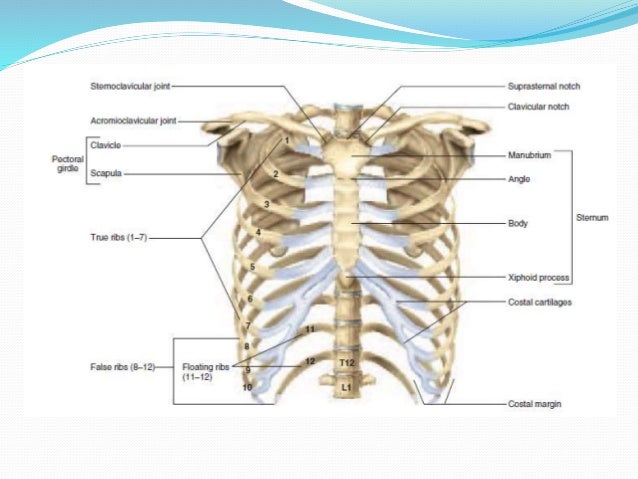
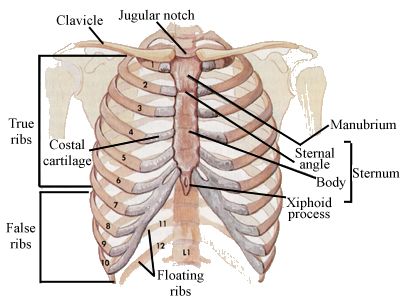
It consists of the 12 pairs of ribs with their costal cartilages and the sternum figure 732. It is supported by the vertical sternum or breastbone anteriorly and the 12 thoracic vertebrae posteriorly. It is the most superior asp this is a division.
The ribs are anchored posteriorly to the 12 thoracic vertebrae t1t12. The ribs are anchored posteriorly to the 12 thoracic vertebrae t1t12. The thoracic cage protects the heart and lungs.
The sternum consists of the manubrium body and xiphoid process. There are some other muscles that do not comprise the thoracic wall but do attach to it. It is formed by the vertebral column ribs and sternum and encloses the heart and lungs.
There are five muscles that make up the thoracic cage. These include the pectoralis major minor serratus anterior and the scalene muscles. Solutions the thoracic cage is formed by the 12 pairs of ribs with their costal cartilages and the sternum.
The thoracic cage protects the heart and lungs. The ribs are anchored posteriorly to the 12 thoracic vertebrae t1t12. It consists of the 12 pairs of ribs with their costal cartilages and the sternum figure 1.
The intercostals external internal and innermost subcostals and transversus thoracis. In humans the rib cage also known as the thoracic cage is a bony and cartilaginous structure which surrounds the thoracic cavity and supports the pectoral girdle shoulder girdle to form the core portion of the human skeleton. Body manubrium what group of bones are shown.
A typical rib is a flattened curved bone. It consists of the 12 pairs of ribs with their costal cartilages and the sternum link. Human anatomy education 73705 views.
Ribs and costal cartilages duration. The thoracic cage rib cage is the skeleton of the thoracic wall. It is formed by the 12 thoracic vertebrae 12 pairs of ribs and associated costal cartilages and the sternum.
These muscles act to change the volume of the thoracic cavity during respiration. Ribs are classified based on if and how their costal cartilages attach to the. The thoracic cage rib cage forms the thorax chest portion of the body.
 Rib Cage Anatomy Human Rib Cage Info And Pictures Rib
 Thoracic Cage Stock Photos Thoracic Cage Stock Images Alamy
 Anterior Thoracic Cage Anatomy
 Human Rib Cage Images Stock Photos Vectors Shutterstock
 Thoracic Cage Injuries
 Anatomy Ch 7 Exam 2 Thoracic Cage Anterior View Diagram
 7 4 The Thoracic Cage Anatomy And Physiology
 Rib Cage And Human Stock Photos Rib Cage And Human Stock
 Anatomy Human Rib Cage
 Unit Iv
 Human Medecine Thoracic Cage Distortion Anatomy Of
 Thoracic Cage
 Thoracic Cage Anatomy Body Human Clipart K60472664
 Ribs Anatomy Types Ossification Clinical Significance
 The Thoracic Cage Scientist Cindy
 Human Skeleton Showing A Transparent Lung With Surrounding Rib Cage D1243 6 012
 Ribs And Sternum Rib Cage Anatomy And Function
-165A5D083AD5D5F81F1-thumb400.jpg) Labeling Thoracic Cage And Vertebrae Human Anatomy 160
Labeling Thoracic Cage And Vertebrae Human Anatomy 160
 Thoracic Cage Is Made Up Of Bones And Cartilage Along It Consists

 Rib Cage Wikipedia

 Thoracic Cage
 Unit Iv
![]() Thoracic Muscles Attachments Actions Teachmeanatomy
Thoracic Muscles Attachments Actions Teachmeanatomy
 Anatomy Of Skeleton Of Thoracic Cage Anterior View Diagram
 Thoracic Cage Anatomy Body Human
 Rib Cage Bones Human Skeletal System Anatomy Vector Illustration
 Rib Cage


Posting Komentar
Posting Komentar