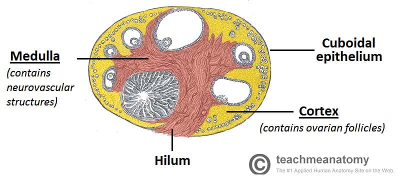
The surface layer of the ovary is formed by simple cuboidal. They also generate the female sex hormones estrogen and progesterone.
 Anatomy Of Female Genital Tract
In width and about 8 mm.
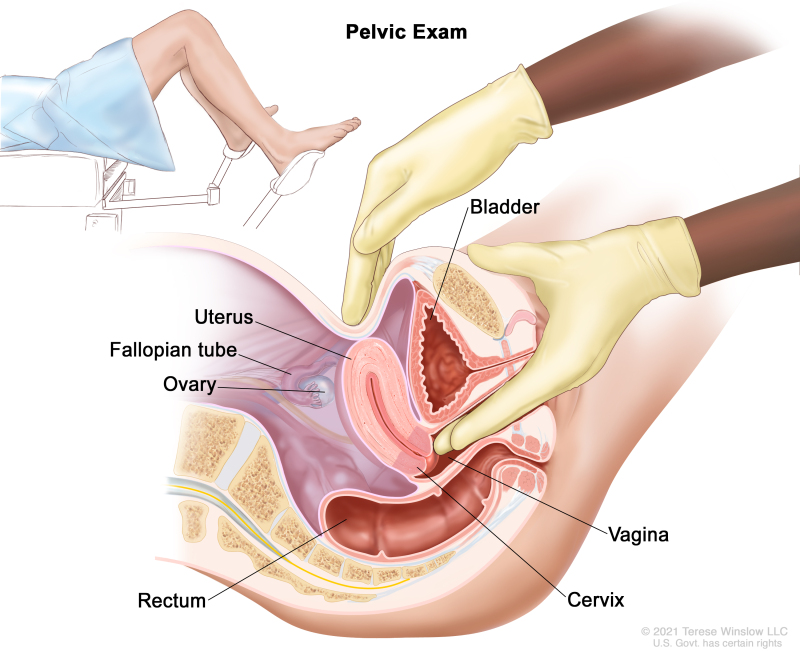
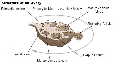
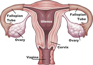
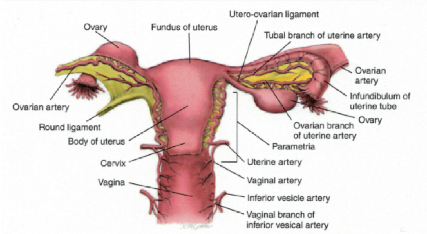
Anatomy of ovary. The ovaries are a pair of oval structures about 15 inches 4 cm long on either side of the uterus in the pelvic cavity fig. In thickness and weigh from 2 to 35 gm. Each month the ovaries go through a series of stages depending on stimulation by.
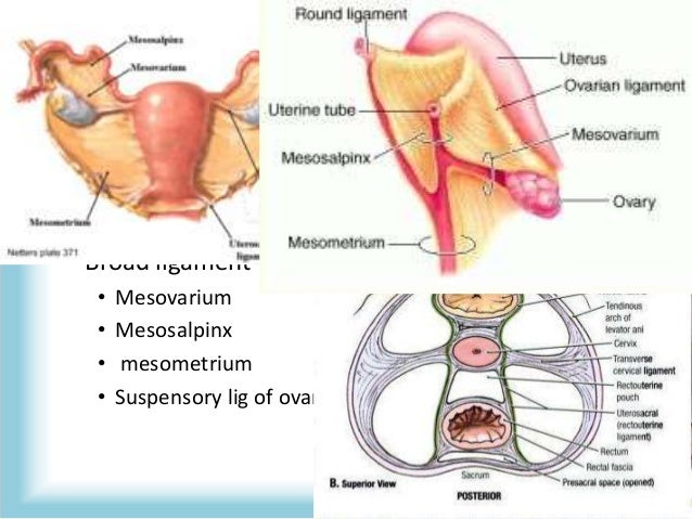
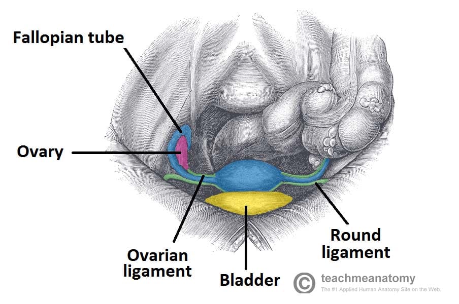
Genital ridge is covered by germinal epithelium previous coelomic epithelium. Connective tissue layer covering the ovarian cortex. Suspensory ligament of ovary fold of peritoneum extending from the mesovarium to.
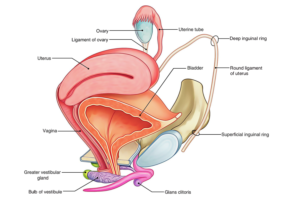
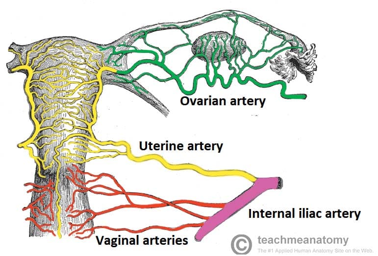
In length 2 cm. Blood supply to the ovary is via the ovarian artery. The ovarian ligament extends from the medial side of an ovary to the uter ine wall and the broad ligament is a fold of the peri toneum that covers the ovaries.
Several paired ligaments support the ovaries. Anatomy of ovaries development. Since the anatomy and function of the ovary vary considerably at different stages in a womans life these aspects will be considered during adulthood childhood and after the menopause.
The ovaries components of the ovary. There is an ovary from latin ovarium meaning egg nut found on each side of the body. The main arterial supply to the ovary is via the paired ovarian.
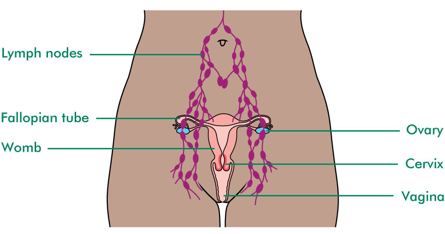
Abstract this chapter deals with the normal macroscopic microscopic and ultrastructural morphology of the human ovary and its hormonal function. Blood supply nerve supply and lymph drainage. They produce the ova eggs that when fertilized will develop into a fetus.
Ovary anatomy the ovaries are female reproductive organs that are akin to the testes in men. The ovaries are of a grayish pink color and present either a smooth or a puckered uneven surface. When a female infant is born her ovaries will contain approximately 400000 egg producing follicles and for the most part her body will not produce anymore follicles for the rest of her life.
Stromal cells resemble fibroblasts. Gross anatomy the female cycle. Formation of primary ovary in female foetus takes place by 10th week coelomic epithelium on medial side of the mesonephros becomes thickened to form genital ridge the site where ovary develops.
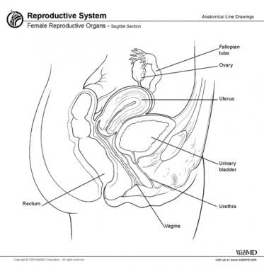
Ovary anatomy physiology introduction. The ovary is the female gonad homologous to the male testes. When released this travels down the fallopian tube into the uterus where it may become fertilized by a sperm.
Anatomy of human ovaries the ovaries develop along with other organs in the womb before birth. They are each about 4 cm. These ligaments help keep the ovaries in place.
In the dog and cat ovaries do not migrate in development. The ovary is an organ found in the female reproductive system that produces an ovum.
 Seer Training Ovaries
 Ovary Anatomy Gross Anatomy Microscopic Anatomy Natural
 Ovarian Cysts Causes Symptoms Treatment Live Science
 Ovarian Epithelial Fallopian Tube And Primary Peritoneal
 The Ovaries Structure Ligaments Vascular Supply Function
 Science Source Ovary Anatomy And Ovulation Cycle Labeled
 File Anatomy Of The Ovaries Jpg Wikimedia Commons
 Easy Notes On Ovaries Learn In Just 4 Minutes Earth S Lab
 The Ovaries Structure Ligaments Vascular Supply Function
 Female Reproductive System Ovary
Anatomy Of The Uterus Uterine Tubes And Ovary Anat10110
 Ovary Model Female Reproductive System Anatomy
 Science Source Ovary Anatomy And Ovulation Cycle Illustration
 Reproductive Anatomy And Physiology Sciencedirect
![]() Ovaries Anatomy And Embryology Kenhub
Ovaries Anatomy And Embryology Kenhub
 2 3 Ovarian Cycle And Female Reproductive Anatomy Diagram
Ovarian Fallopian Tube And Primary Peritoneal Cancer
 Ovary Of Female Female Reproductive System Anatomy
 The Ovaries Structure Ligaments Vascular Supply Function

 The Ovaries Fallopian Tubes And Peritoneum Macmillan
 Pathological Female Uterus And Ovary Medical Anatomy Model
 Female Reproductive System Human Anatomy Uterus And Ovaries
/male_female_gonads-58811e985f9b58bdb3e3dfe9.jpg) Male And Female Gonads Testes And Ovaries
Male And Female Gonads Testes And Ovaries


Posting Komentar
Posting Komentar