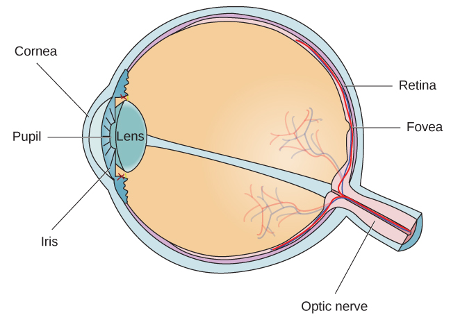
The eye uses the retina to produce an image whilst the camera uses film. It changes shape to help the eye focus to see objects clearly at near.
 Reston Eye Anatomy Eye Anatomy In Reston Eye Anatomy 20194
Anatomy of the eye.

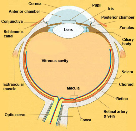
The anatomy of the eye. The lens of the eye is located directly behind the pupil. The diagrams below show cross sections of the human eyeball. Anatomy of the eye.
Anatomy of the eye the eye can be compared to a camera. Vision is our window to the outside world. Extraocular muscles eye anatomy duration.
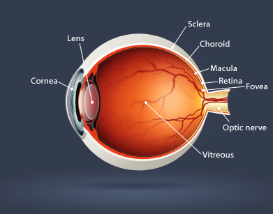
Picture of eye anatomy detail. This article explores the anatomy of the eye looking at the different structures of the human eye and their function. They both focus the incoming light with the help of a lens.
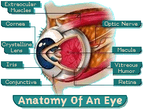
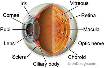
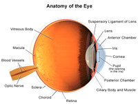
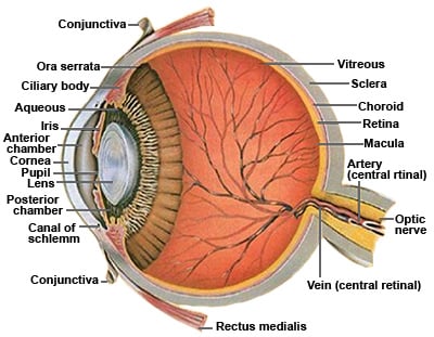
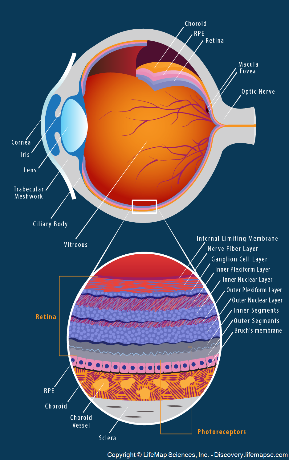
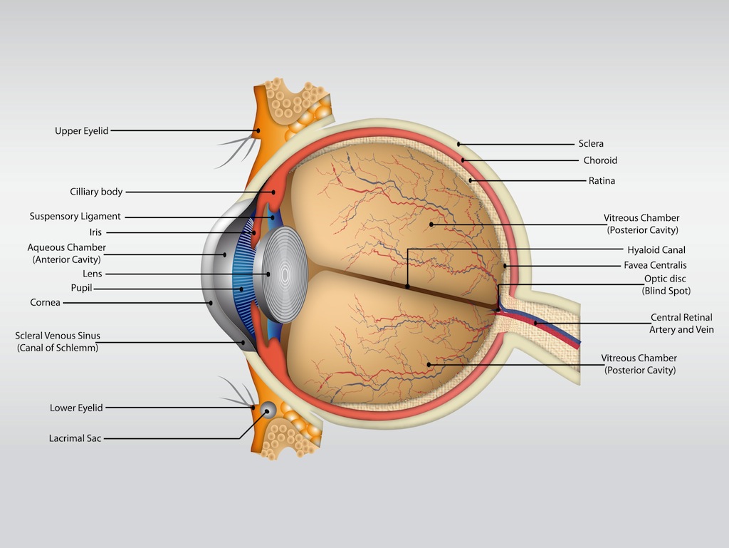
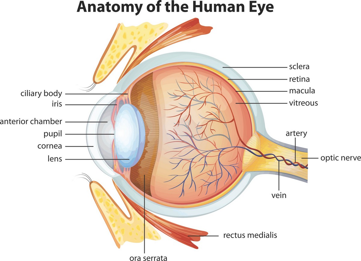
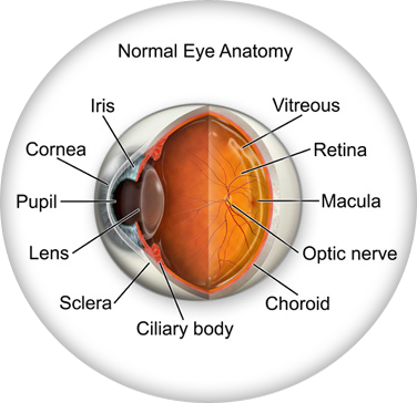
Nerve signals that contain visual information are transmitted through the optic nerve to the brain. An area of tissue in the eye located around the base of the cornea near the ciliary body and is responsible for draining the aqueous humor from the eye via the anterior chamber sclera the white of the eye is the opaque fibrous protective outer layer of the eye containing collagen and elastic fiber. The inside lining of the eye is covered by special light sensing cells that are collectively called the retina.
Clear front window of the eye that transmits and focuses light into the eye. And when there is low light the iris opens up the pupil to let in more light. Unsubscribe from armando hasudungan.
Anatomy of the eye. Anatomy eye overview armando hasudungan. The eye is our organ of sight.
The eye is the organ responsible for vision. When there is bright light the iris closes the pupil to let in less light. It converts light into electrical impulses.
The eye is surrounded by the orbital bones and is cushioned by pads of fat within the orbital socket. Lens focuses light rays onto the retina. The eye has a number of components which include but are not limited to the cornea iris pupil lens retina macula optic nerve choroid and vitreous.
They both gather light and transform it into a picture that we can interpret. Extraocular muscles help move the eye in different directions. A closer look at the parts of the eye by liz segre when surveyed about the five senses sight hearing taste smell and touch people consistently report that their eyesight is the mode of perception they value and fear losing most.
Behind the eye your optic nerve carries. Iris the colored part of the eye which helps regulate the amount of light entering the eye. The lens bends light coming into the eye to help focus it on the retina.
 Special Senses Vision Anatomy And Physiology I
 Muscle Identification Eye Anatomy Human Anatomy Anatomy
 Amazing Anatomy Parts Of The Eye Defined Michigan Eye
 Eye Anatomy Glaucoma Research Foundation
 Blind Spot Anatomy Britannica
![]() Blood Vessels And Nerves Of The Eye Anatomy Kenhub
Blood Vessels And Nerves Of The Eye Anatomy Kenhub

 Choroid Wikipedia
 Eye Anatomy Ocular Anatomy Vision Conditions Problems

 Eye Anatomy Definitions
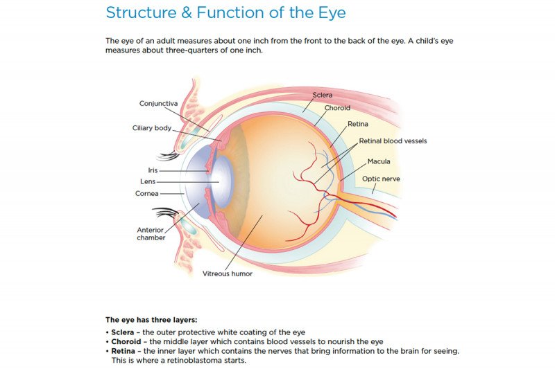 Retinoblastoma Anatomy Of The Eye Memorial Sloan
 Vision Introduction To Psychology
 Eye Structure And Function In Cats Cat Owners Merck
 Eye Anatomy Vector Vector Art Graphics Freevector Com
 Vision And The Eye S Anatomy Healthengine Blog
 Anatomy Of The Eye Children S Wisconsin
 Eye It S Nice To Meet You The Anatomy Of The Eye And How
 Anatomy Of The Human Eye Visual Acuity Light Perception
 Eye Anatomy Highlands Ranch Co


Posting Komentar
Posting Komentar