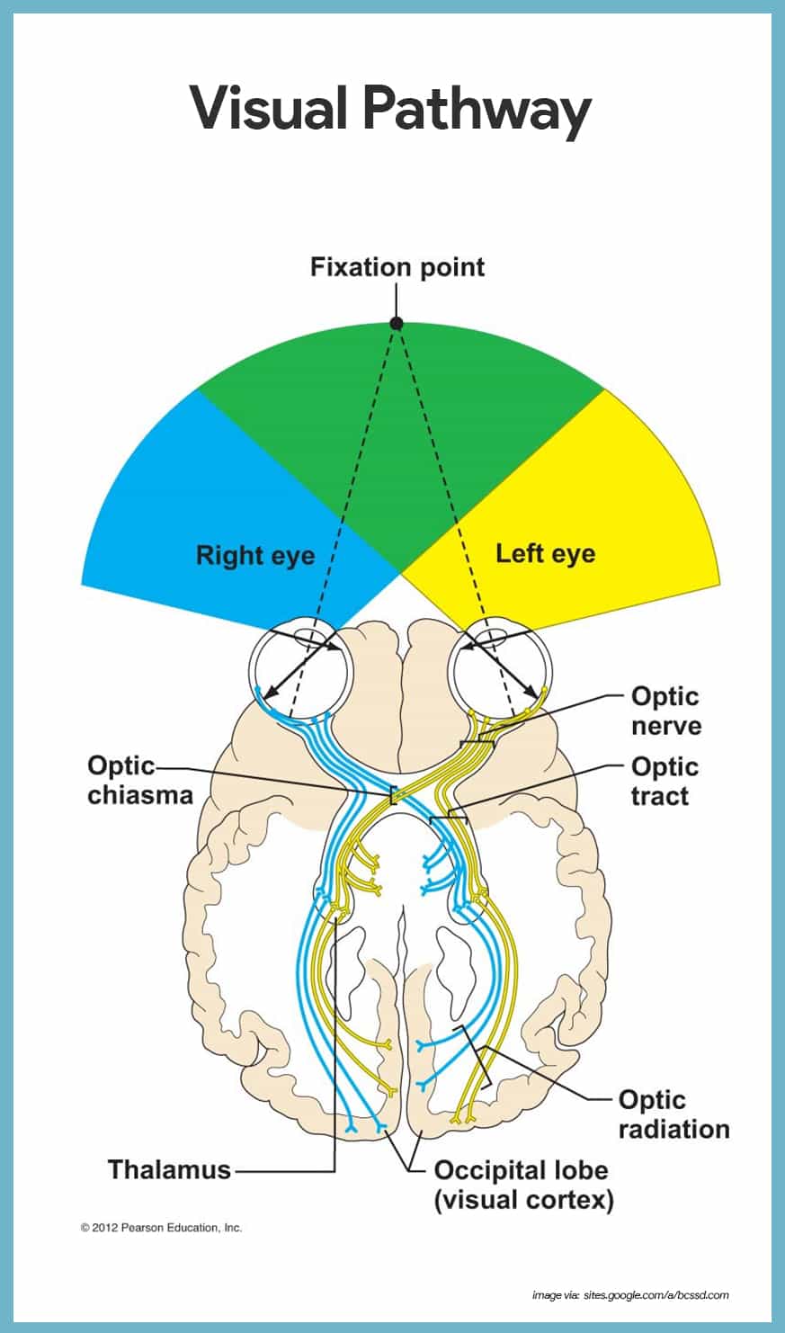
Nerve signals that contain visual information are transmitted through the optic nerve to the brain. The human eye is an organ that reacts to light and has several purposesthe eye is a complex structure with layers lens muscles receptors that is surrounded by many boneswatch various parts.
 Eye Anatomy And Vision Course Hero
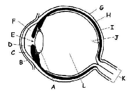
Eye ball structures 1 fibrous tunic 2 vascular tunic 3 nervous tunic 3.

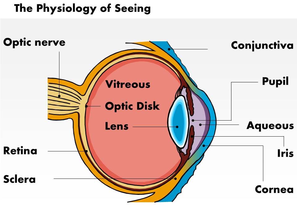
The eye anatomy and physiology. In a normal eyeball after exiting the back of the lens the light rays pass through the vitreous a clear jelly like substance that fills the globe of the eyeball. It has two types of muscles. The vitreous humor helps the eye hold its spherical shape.
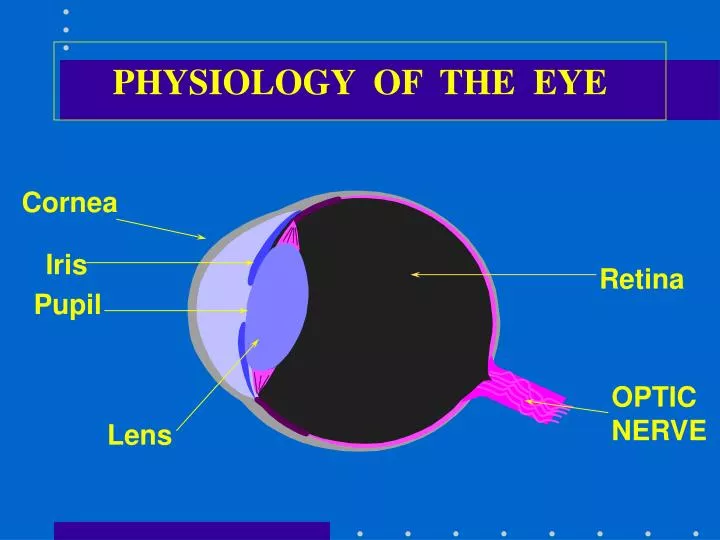
Physiology of the eye 1. Anatomy of the eye. Development anatomy and physiology of the eye the word perspective comes from the latin per through and specere look at.
By bahaa halwany department of ophthalmology medicals international 2. Last week we discussed vision from a historical perspective in order to understand how johannes kepler rené descartes and bishop berkeley discovered the importance of the mind in effecting vision. The eye is the organ of the visual system and responsible for processing visual detail.
Extraocular muscles help move the eye in different directions. Interior of the ball 1 anterior cavity 2 vitreous chamber 3 lens b. They have more rods.
Finally the light rays land and come to a sharp focusing point on the retina. The eye is surrounded by the orbital bones and is cushioned by pads of fat within the orbital socket. A tapetum is a mirror type membrane that is reflective on the back of the eye activating the rod gives better night vision vitreous humor posterior portion of eye filled with jelly like substance.
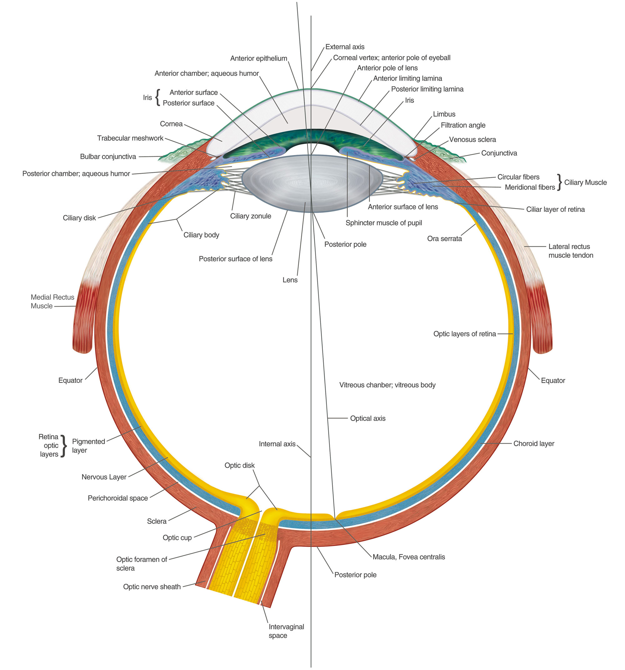
Circular and radial muscle. Anatomy and physiology of the eye 1. The eye is made up of a outer fibrous layer a middle vascular layer and an inner retinal layer where sensory.
Anatomy of the eye. It is a complex optical system which collects light and converts it to electrochemical impulses in neurons. Anatomy and physiology of the ear.
It has small circular opening called pupil. It is muscular pigmented and opaque diaphragm which hangs in the eye ball in front of lens. Conjunctiva cornea iris lens macula retina optic nerve vitreous and extraocular muscles.
Anatomy physiology pathology of the human eye included are descriptions functions and problems of the major structures of the human eye.
 The Anatomy And Physiology Of Animals Special Senses

 Anatomy Physiology 1 Final Exam
 Visual Pathway Human Anatomy Physiology Eye Anatomy
 Amazon Com Semtomn Gaming Mouse Pad Blue Components Of
 Arrangement Of The Extraocular Muscles From Anatomy
Eye Structure And Function Anatomy And Physiology Flashcards
 Simple Eye Anatomy And Physiology Liquid Conscience
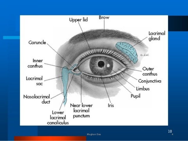 Anatomy And Physiology Of Eye By Maghan Das
 A P 2 Lab Test Eye Ear Anatomy Physiology 123 With
 Special Senses Anatomy And Physiology Nurseslabs
 Alternative Healing Basic Anatomy And Physiology Of The
 Special Senses Vision Anatomy And Physiology I

 Ppt Physiology Of The Eye Powerpoint Presentation Free
 Eye Anatomy And Physiology A Complete Detail With Images
 Anatomy And Physiology Sensory System The Eye
 0514 Physiology Of Seeing Eye Anatomy Medical Images For

 Worksheet With Answer For Structure Of Human Eye Eye
 Anatomy And Physiology Of The Human Eye Effects Of
 The Eyes Canadian Cancer Society
 Special Senses Anatomy And Physiology Nurseslabs
 Eye Anatomy Glaucoma Research Foundation


Posting Komentar
Posting Komentar