This lecture is a part of basic radiologic anatomy series. As myelin is a fatty substance it is of relatively low density compared to the cellular grey matter.
 Normal Anatomy Of The Brain On Ct And Mri With A Few Normal
Anatomy of the head on a cranial ct scan.

Anatomy of brain ct scan. Anatomy of the head on a cranial ct scan. The app has a full body ct scan and uses color coded pins to label the anatomic structures. The brain consists of grey and white matter structures which are differentiated on ct by differences in density.
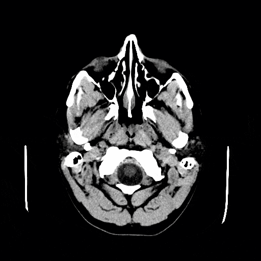
Ct scan provides a 3d display of the intracranial anatomy built up from a vertical series of transverse axial tomograms each tomogram represents a horizontal slice through the patients head. It is great for learning general anatomy or showing patients a normal ct scan for comparison. 6 frontal bone 27 occipital bone 32 optic nerve 43 frontal sinus 45 sigmoid sinus 46 internal carotid artery.
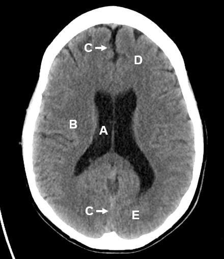
This means that the right side of the brain is on the left side of the viewer. Anatomy ct axial brain anatomy ct axial brain form no 1. White matter has a high content of myelinated axons.
Brain bones of cranium sinuses of the face. Grey matter contains relatively few axons and a higher number of cell bodies. Brain and face ct.
Ct images of the brain are conventionally viewed from below as if looking up into the top of the head. Learn ct scan learn the diagnosis of ct and methods of computed tomography. Ct scans are created using a series of x rays which are a form of radiation on the electromagnetic spectrum.
They lie on the ventricular surface of the hippocampus and become the fimbria of the fornix medially. Brain bones of cranium sinuses of the face. Anatomy ct axial brain form no 19.
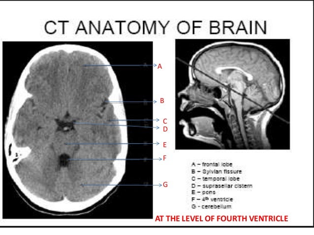
The lecture discussing the basic ct anatomy of the brain. Head ct anatomy normal anatomy 1. The anterior part of the head is at the top of the image.
Amygdala on ct and mr images the amygdala is a large region of gray matter contiguous with the uncus of the medial temporal lobe and the most anterior portion of the hippocampus the pes hippocampi. Ct anatomy is a useful radiology app that helps educate the user on normal human anatomy seen on the ct. The scanner emits x rays towards the patient from a variety of angles and the detectors in the scanner measure the difference between the x rays that are absorbed by the body and x rays that are transmitted through the body.
 Ct Scans Interpretation Principles Basics Teachmeanatomy
Mri Ct And High Resolution Macro Anatomical Images With
 Radiology Basics Head Anatomy

 Basic Anatomy Of Ct Brain Hku E Learning Platform In
 Normal Anatomy Radiology Key
 Ct Scans Interpretation Principles Basics Teachmeanatomy

 Figure 69 3 From How To Read A Head Ct Scan Semantic Scholar
 Ct Scan Of Head And Neck
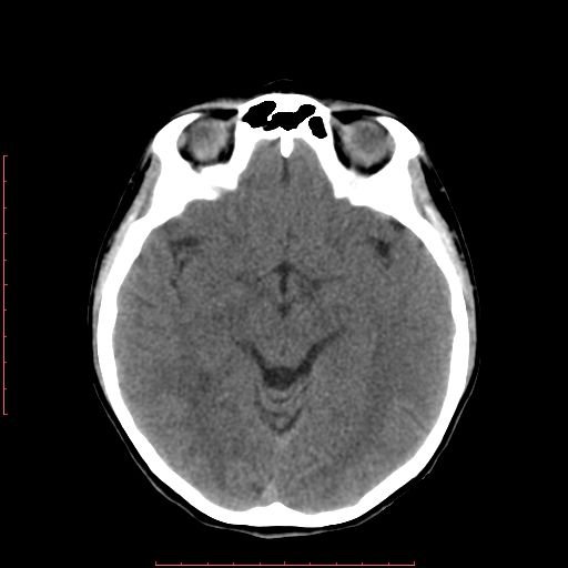
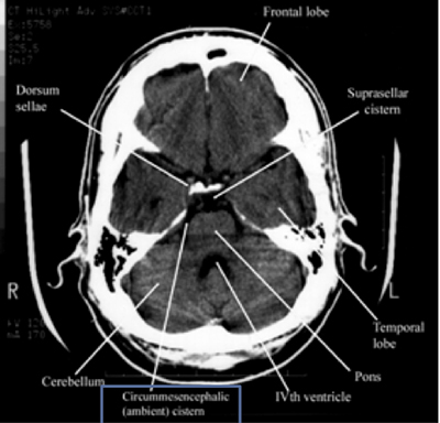
 Normal Ct Brain
 Radiology Basics Head Anatomy
Normal Head Ct
 Ct Head Interpretation Radiology Geeky Medics
 Cns Anatomy Springerlink
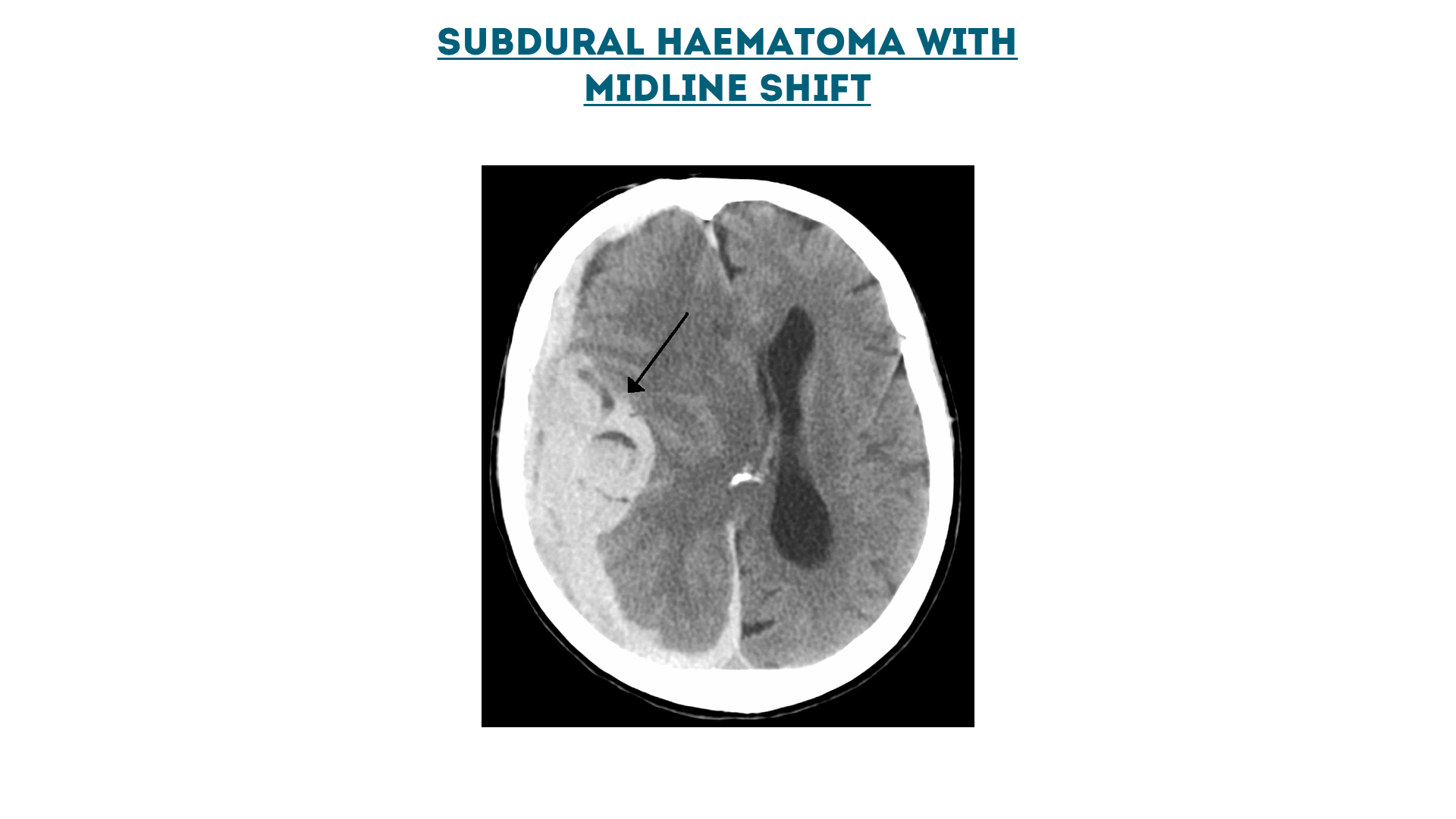
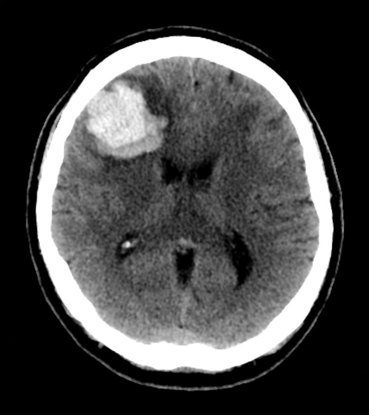
 Ct Brain Hemorrhage Startradiology
 Brain Ct Anatomy Cerebral Lobes Ventricles Brain
 Ct Brain Hemorrhage Startradiology
 The Radiology Assistant Brain Anatomy

 Ct Axial Anatomy
 The Radiology Assistant Brain Anatomy
 The Radiology Assistant Brain Anatomy
Normal Head Ct
 How To Read A Head Ct Emergency Medicine Newyork
 Test Yourself Ct Brain Quiz 2
Hounsfield Scale An Overview Sciencedirect Topics
 Figure 69 5 From How To Read A Head Ct Scan Semantic Scholar
![]() Medical Imaging And Radiological Anatomy X Ray Ct Mri
Medical Imaging And Radiological Anatomy X Ray Ct Mri
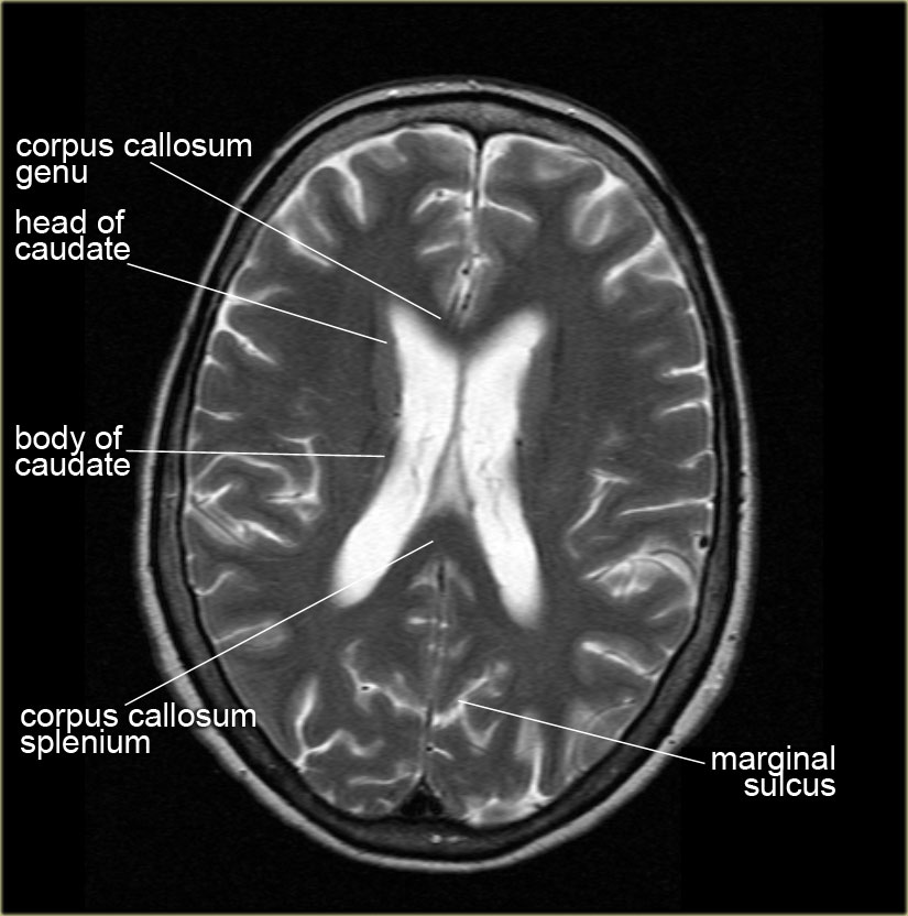
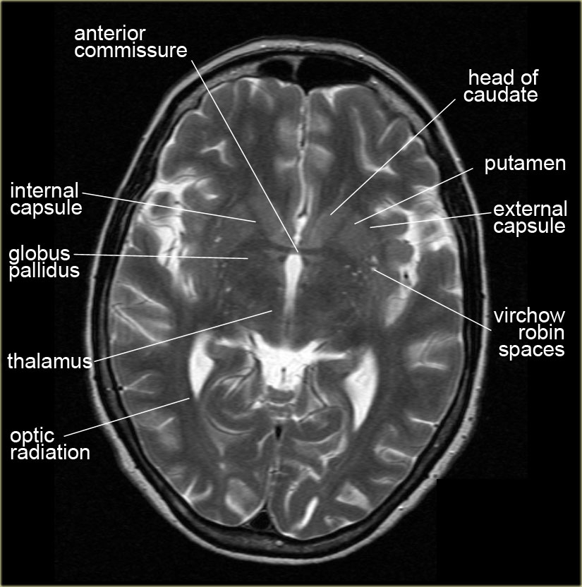
 Mri Anatomy Free Mri Axial Brain Anatomy
 Normal Ct Brain Radiology Case Radiopaedia Org


Posting Komentar
Posting Komentar