The outer layer of the eye consists of 8 eye parts. The white part of the eye is a tough outer layer called the sclera.
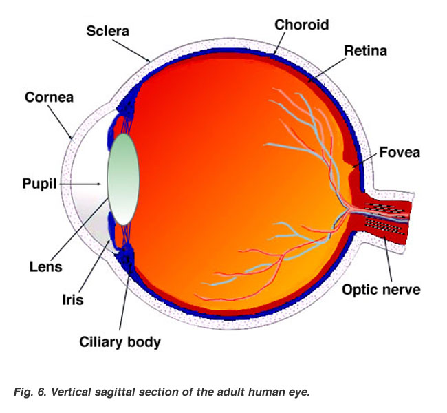
Human eye is spherical about 25 cm in diameter.
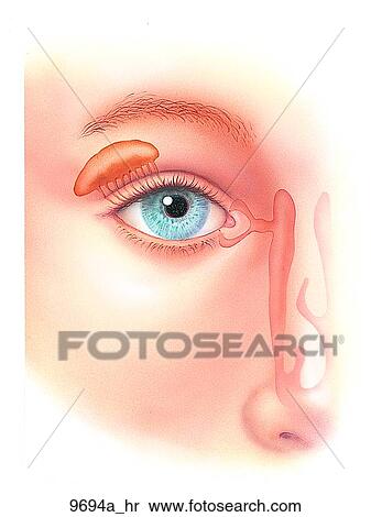
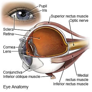
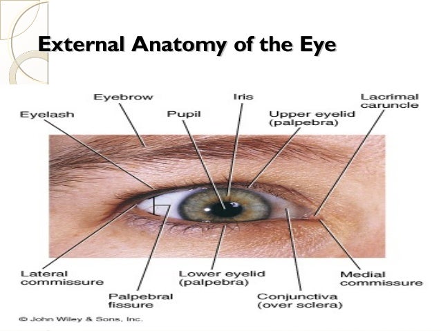
Anatomy of the outer eye. It lies in front of the iris the coloured part of the eye. Extraocular muscles help move the eye in different directions. The cornea is like a window into the eye.
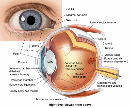
The anatomy of the eye includes the cornea pupil lens sclera conjunctiva and more. The cornea is the outer covering of the eye. Iris the colored part of the eye which helps regulate the amount of light entering the eye.
The colored part of the eye is called the iris. The outer eye when looking at the outside of the eye several structures are readily available for viewing. Nerve signals that contain visual information are transmitted through the optic nerve to the brain.
The outer layer contains the sclera the white of the eye and the cornea the clear dome at the front of the eye. Anatomy of the eye. The sclera protects the inside of the eye and helps the eye keep its structure.
It is situated on an orbit of skull and is supplied by optic nerve. Anatomy of the eye the ottawa hospital. This dome shaped layer protects your eye from elements that could cause damage to the inner parts of the eye.
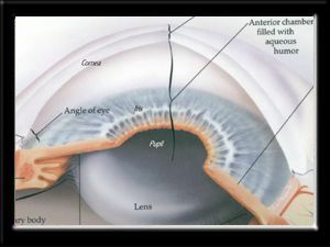
And when there is low light the iris opens up the pupil to let in more light. The cornea is like a window it helps to focus light onto the retina. The iris acts like the shutter of a camera regulating the amount of light that enters.
The eye is surrounded by the orbital bones and is cushioned by pads of fat within the orbital socket. There are many parts of the eye. When there is bright light the iris closes the pupil to let in less light.
There are 6 sets of muscles attached to outer surface of eye ball which helps to rotate it in different direction. Eye part 1 cornea. Anatomy of the eye.
The eye is the photo receptor organ. Anatomy parts and structure. Lens focuses light rays onto the retina.
When light passes through the eye the cornea refracts the light rays in a way so that it can land directly on the retina. The transparent dome like structure that is covering the iris and the pupil.
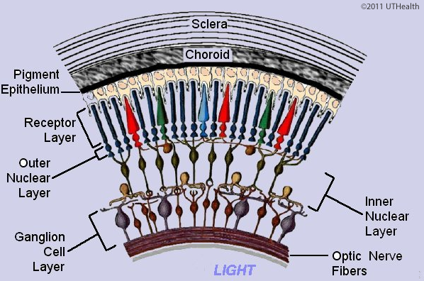
Neuroanatomy Online Lab 7 Visual System Microscopic
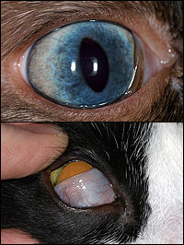 Why Do Cats Have An Inner Eyelid As Well As Outer Ones
 Human Eye Ball Anatomy Physiology Diagram

 Human Eye Movements Of The Eyes Britannica
 Ucsd S Practical Guide To Clinical Medicine
 The Eye And Vision
:max_bytes(150000):strip_icc()/GettyImages-695204442-b9320f82932c49bcac765167b95f4af6.jpg) Structure And Function Of The Human Eye
Structure And Function Of The Human Eye
 Eye Anatomy Glaucoma Research Foundation
 Cow S Eye Dissection Eye Diagram
 Right Eye Outer Anatomy Unlabeled Stock Illustration
 Iris And Uvea Of The Eye Allaboutvision Com
 Human Eye Ball Anatomy Physiology Diagram
 Anatomy Of The Eye The Ottawa Hospital
 Eye Anatomy Definitions
 Anatomy Of The Eye
 Eye Anatomy Central Florida Retina
 Corneal Flash Burns What You Need To Know
 Special Senses Vision Anatomy And Physiology I
 Human Eye Anatomy Parts Of The Eye And Structure Of The
Parts Of The Eye American Academy Of Ophthalmology
 Ppt On Eye Anatomy
 Ucsd S Practical Guide To Clinical Medicine
 Retinoblastoma Symptoms And Causes Mayo Clinic

 Eye Anatomy Detail Picture Image On Medicinenet Com


Posting Komentar
Posting Komentar