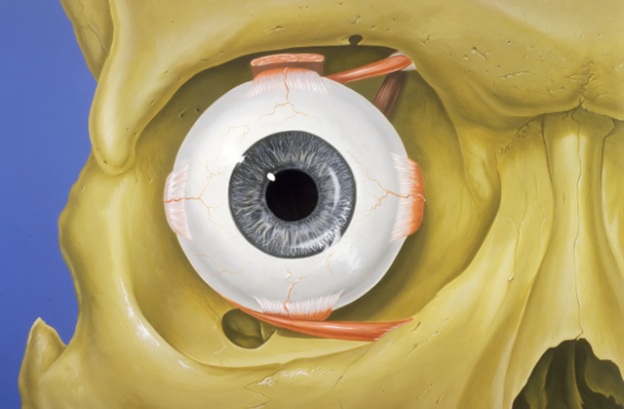
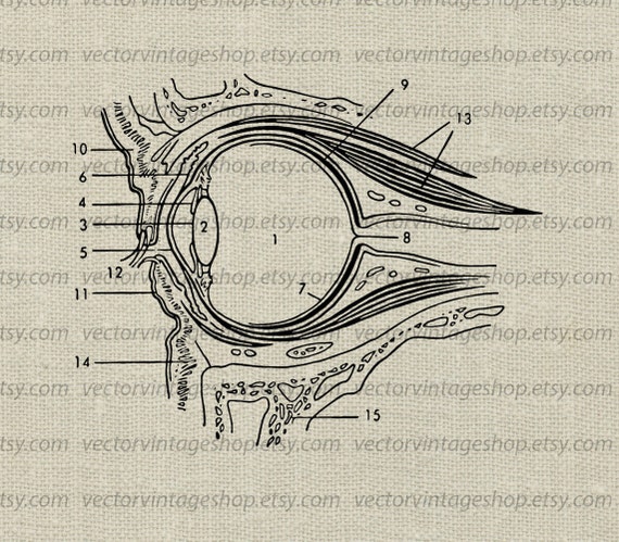
The diagrams below show cross sections of the human eyeball. Eye anatomy bones of the orbit.

As a sense organ the eye allows vision.

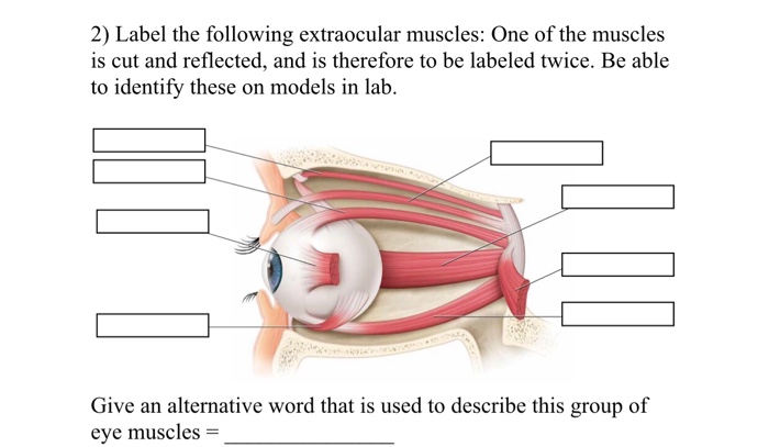
Eye muscles anatomy. The actions of the six muscles responsible for eye movement depend on the position of the eye at the time of muscle contraction. The primary action of the superior oblique muscle is intorsion or internal rotation the secondary action is depression. The bony orbit is made out of seven bones which include the maxilla.
The other four muscles move the eye up down and at an angle. The eye is the organ responsible for vision. They can be divided into two groups.
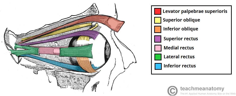
Muscles of eye movement. The extraocular muscles are the six muscles that control movement of the eye and one muscle that controls eyelid elevation levator palpebrae. Would you like to see your own top scores for this anatomy game in this space.
There are six muscles involved in the control of the eyeball itself. This article explores the anatomy of the eye looking at the different structures of the human eye and their function. There is also minor function in lateral rotation.
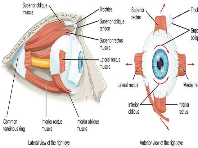
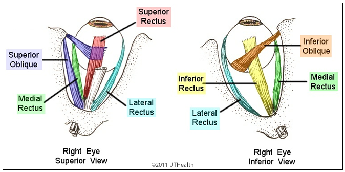
These muscles are named the superior rectus inferior rectus lateral rectus medial rectus superior oblique and inferior oblique. The two oblique muscles of the eye are responsible for the rotation of the eye and assist the rectus muscles in their movements. There are four recti muscles.
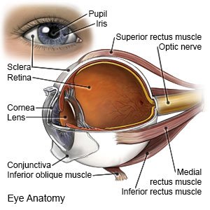
Anatomy of the eye. Eye muscle anatomy there are six extraocular muscles that move the globe eyeball. These muscles characteristically originate from the common tendinous ring.
There are six eye muscles that control eye movement. The superior oblique muscle rotates the eye medially and abducts it when the eye if facing forward while the inferior oblique rotates the eye laterally and adducts it. Use this anatomy quiz to learn to locate its components.
Vision is our window to the outside world. Six extraocular muscles. One muscle moves the eye to the right and one muscle moves the eye to the left.
The lacrimal gland is a part of the lacrimal apparatus. The four recti muscles and the two oblique muscles. The human eye is an organ which reacts to light and pressure.
The eyelids are soft tissue structures that cover and protect. Its antagonist is the lateral rectus muscle that abducts the eye allowing it to look laterally or away from the bodys midline. Superior rectus inferior rectus medial rectus and lateral rectus.
The eye anatomy games.
 Eye Muscles Medlineplus Medical Encyclopedia Image
 Extra Ocular Muscles Anatomy
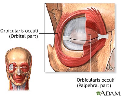
 Which Cheek Nose And Outer Eye Muscles Are Involved In
 Extraocular Muscles Pic An0002

Anatomy Of The Eye American Association For Pediatric
 Muscle Identification Eye Anatomy Human Anatomy Anatomy
 Neuroanatomy Online Lab 7 Visual System Infranuclear
 Eye Opener Anatomy Muscles Of The Eye
 Extraocular Muscles Basic And Clinical Anatomy Lecturio
 Amazon Com Optic Chiasm And Eye Muscles Watercolor Poster
 The Extraocular Muscles The Eyelid Eye Movement
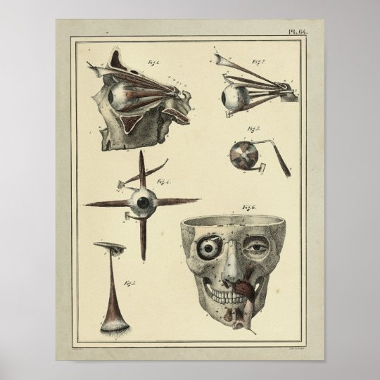 Vintage 1831 Eye Muscles Anatomy Print
 Extrinsic Eye Muscles Diagram Quizlet
 Extrinsic Eye Muscles Purposegames
 Extrinsic Eye Muscles At University Of Wisconsin Lacrosse
 Strabismus In Children What You Need To Know
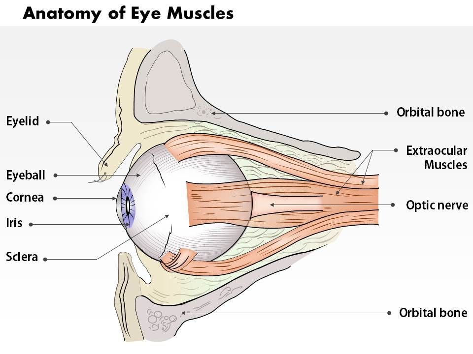 0514 Anatomy Of Eye Muscles Medical Images For Powerpoint
 Llustration Of The Extraocular Muscle Anatomy Orientation
 Eye Anatomy Clipart Eye Muscles Diagram Medical Vector Clip Art Science Vintage Illustration Medical Art Instant Download Commercial Use
 Pediagenosis
 Slide Show A Look Inside Your Eyes Mayo Clinic
 Ophthalmoplegia Eye Disorder Britannica

 Researcher Wants To Unlock The Mysteries Of Strabismus
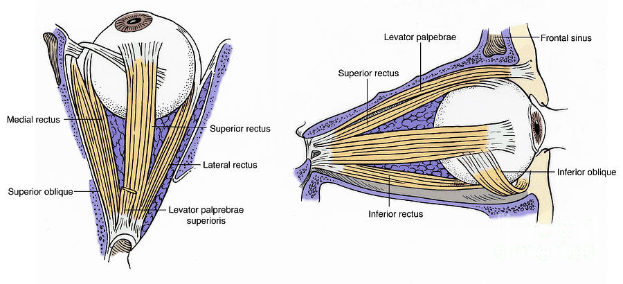 Illustration Of Eye Muscles By Science Source
 Eye Muscles Anatomy Chapter 10 Flashcards Quizlet
 Solved 25 2 The Extrinsic Muscles Of The Eye Inferior Ob
 Muscles Of The Human Eye Stock Illustration Illustration Of


Posting Komentar
Posting Komentar