The maxillary sinuses are shaped like a pyramid and each contain three cavities which point sideways inwards and downwards. In humans the maxilla consists of.
 Superior Maxillary Bone Clipart Etc
The body of the maxilla.
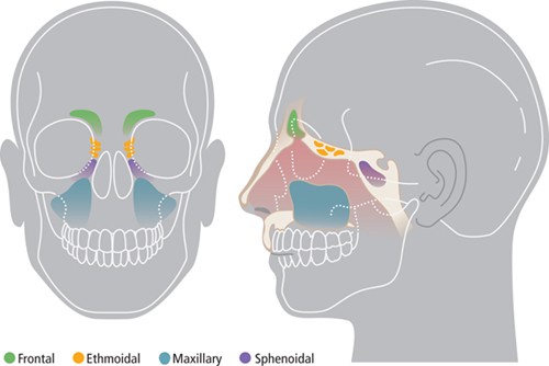
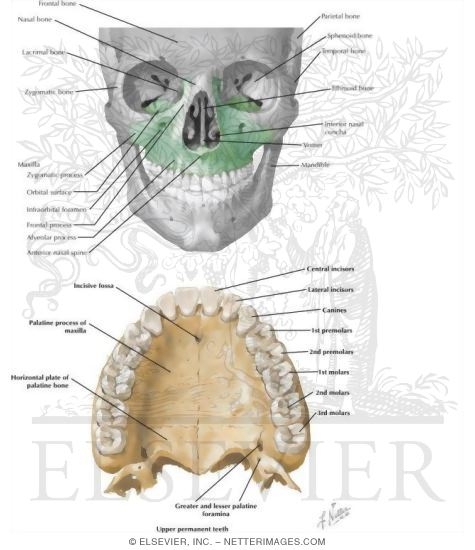
Maxillary anatomy. The two maxillary sinuses are located below the cheeks above the teeth and on the sides of the nose. Each maxilla has four processes frontal zygomatic alveolar and palatine and helps form the orbit roof of the mouth and the lateral walls of the nasal cavity. Forms a large part of nasal floor and anterior three fourths of hard palate.
Anteriorly in the midline articulation of both palatine processes is the incisive canal which transmits the nasopalatine nerve and branches of the greater palatine vessels. The maxillary sinus is the largest of the paranasal sinuses. Maxilla bone anatomy the two maxilla or maxillary bones maxillae plural form the upper jaw l mala jaw.
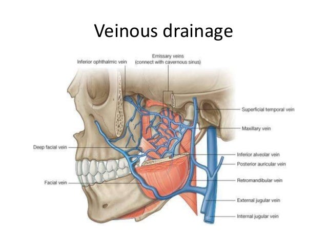
The maxillary bones on each side join in the middle at the intermaxillary suture a fused line that is created by the union of the right and left halves of the maxilla bone. Contains two grooves posterolaterally that transmit the greater palatine vessels and nerves. The maxillary artery is one of the two terminal divisions of the external carotid artery in the head.
Therefore the maxillary artery can be defined as one of the continuations of the external carotid artery and distributes the blood flow to the upper maxilla and lower mandible jaw bones deep facial areas cerebral dura mater and the nasal cavity. The maxilla consists of the body and its four projections. The maxilla forms the upper jaw by fusing together two irregularly shaped bones along the median palatine suture located at the midline of the roof of the mouth.
The second terminal branch is the superficial temporal artery. Three surfaces anterior posterior medial. From a medial view the maxillary hiatus is evident opening into the maxillary sinus that occupies the predominant portion of the body of the maxilla.
Projects medially from lowest part of medial aspect of maxilla. Midline incisive fossa behind.
 External And Internal Root Canal Anatomy Of The First And

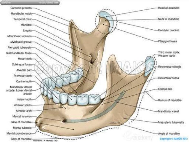
 Facial Bone Anatomy Overview Mandible Maxilla
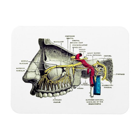
 Maxillary Nerve An Overview Sciencedirect Topics
![]() Maxilla Anatomy Function Clinical Aspects Kenhub
Maxilla Anatomy Function Clinical Aspects Kenhub
 Oral Surgery Ii Part 2 The Maxillary Sinus Antrum And
 Anatomy Of The Maxilla And Mandible Diagram Quizlet
 Maxillary Nerves Human Anatomy Magnet
 Bones Of The Skull Maxilla
 Stock Illustration
 Anatomy Of Maxilla And Mandible
 Complete Denture Anatomical Landmarks My Dental

 Anatomy Branches Of The Maxillary Artery
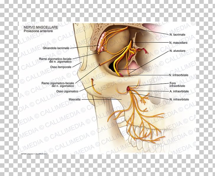 Infraorbital Nerve Maxillary Nerve Zygomatic Nerve Png
 Anatomy Of Maxilla And Mandible
 Maxillary Artery Anatomy Branches
 Maxilla Anatomy Gross Anatomy Anatomy Anatomy Drawing
 Facial Bone Anatomy Overview Mandible Maxilla
 Maxillary Bone
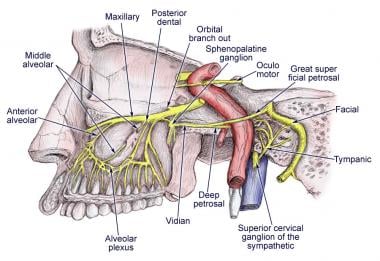
 The Maxillary Nerve Block Anatomy The Maxillary Nerve Exits
 Maxilla Wikipedia
 The Maxillary Division Of The Trigeminal Nerve Cnv2
 Anatomical Sketches Of Leonardo Da Vinci A Dissection Of
 Maxillary Tuberosity Tuberosity Of Maxilla Earth S Lab
 Maxillary Bone
 Maxillary Hiatus Wikiwand
 Maxillary Division Of Trigeminal Nerve V2 Or Vb Maxillary Nerve Anatomy Medical Animations
 Maxilla Wikipedia
Trigeminal Nerve Anatomy Gross Anatomy Branches Of The
 Maxillary Bone Anatomy Diagram Quizlet
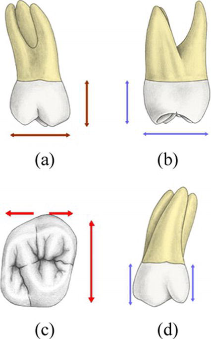
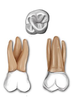 Maxillary First Molar Wikipedia


Posting Komentar
Posting Komentar