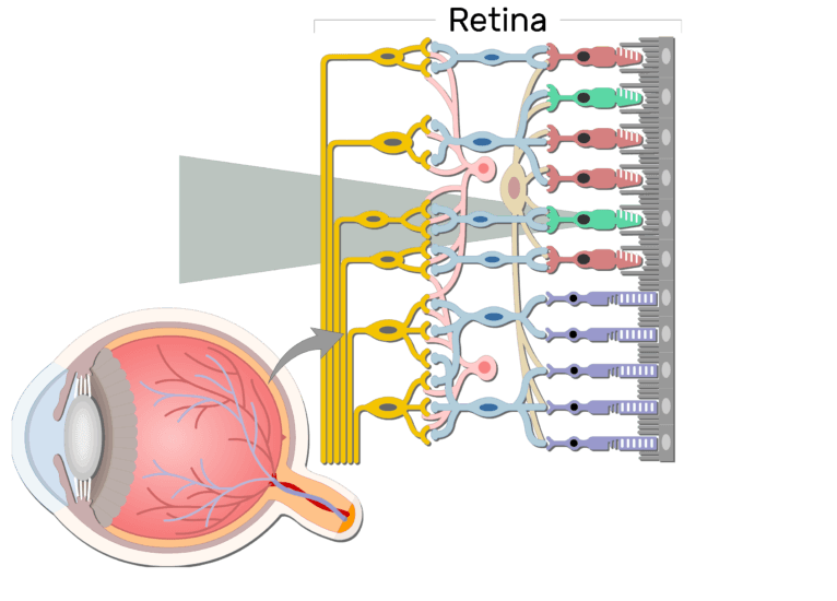
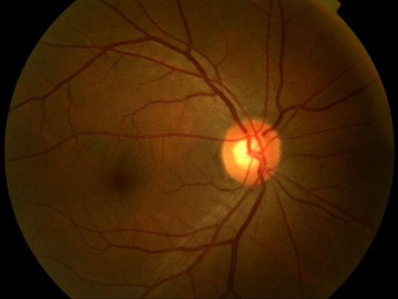
These are essentially light sensitive cells responsible for detecting qualities such as color and light intensity. The red curving structures are blood vessels which enter the retina through the nerve.
 Anatomy Of The Adult Human Eye And Retinal Layers 10 A
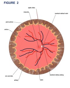
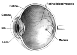
Several parts of the eye are associated with the retina.

Retinal anatomy. The retina processes the information gathered by the photoreceptor cells and sends this information to the brain via the optic nerve. The optic nerve contains the ganglion cell axons running to the brain and additionally incoming blood vessels that open into the retina to vascularize the retinal layers and neurons fig. Read an overview of general eye anatomy to learn how the parts of the eye work together.
The retina is approximately 05 mm thick and lines the back of the eye. Cellular anatomy of the retina. The whitish circle is the nerve that connects the retina to the brain.
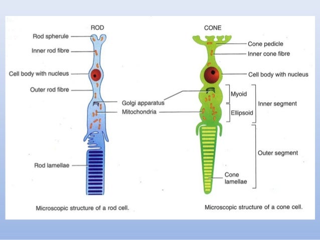
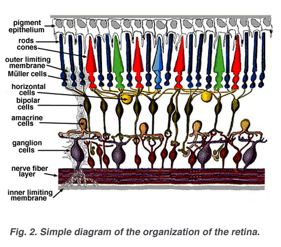
These cells can be divided into a three basic cell types photoreceptor cells neuronal cells and glial cells. The retina processes light through a layer of photoreceptor cells. Refer to this page for comparison with the retinal disease pages.
The optics of the eye create a focused two dimensional image of the visual world on the retina which translates that image into electrical neural impulses to the brain to create visual perception the retina serving a function analogous to that of the film or image sensor in a camera. Photoreceptors rods and cones comprise the inner sensory layer of the retina. This layer senses light and sends signals to the brain so you can see.
This page describes normal retinal anatomy. This fundus photograph shows the normal appearance of the retina. Retina lines the globe inner surface and contains light sensitive neuron s that transmit signals to the optic nerve.
The retina consists of millions of cells packed together in a tightly knit network spread over the surface of the back of the eye. Simple anatomy of the retina by helga kolb. The neural retina consists of several layers of neurons interconnected by synapses and is supported b.
The retina is the innermost light sensitive layer of tissue of the eye of most vertebrates and some molluscs. The retina is the layer of nerve cells lining the back wall inside the eye.
 Retina Wikipedia
 Anatomy Of The Vertebrate Eye The Retina Lining The Inner
 Eye Anatomy Rod Cells And Cone Cells The Arrangement Of Retinal
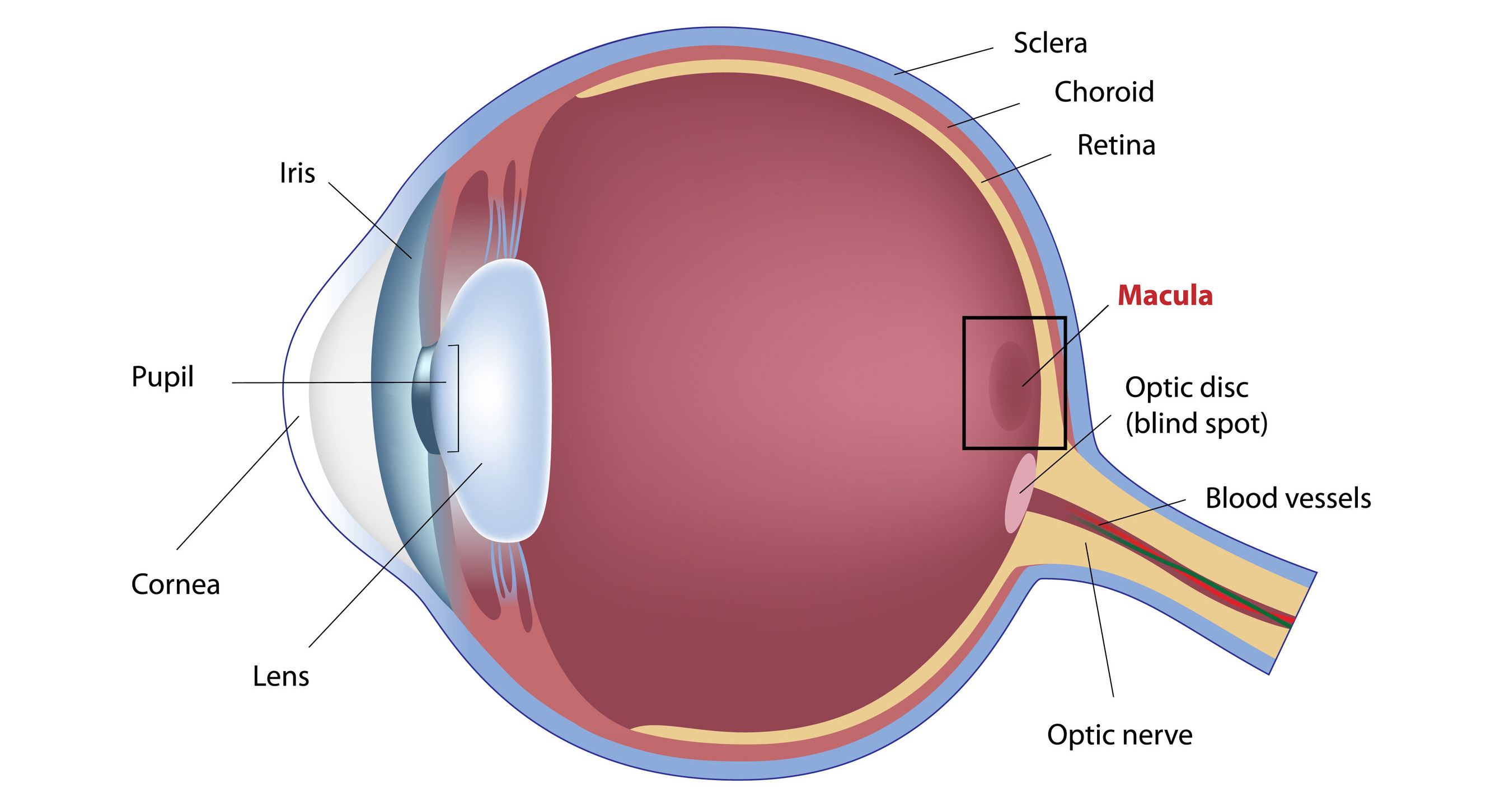 What Is The Macula
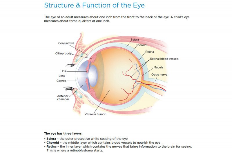 Retinoblastoma Anatomy Of The Eye Memorial Sloan

 Retinopathy Of Prematurity Rop
 Illustration Of Eye Anatomy And Retinal Layers 2 3 A
 Anatomy Of Retina
 Vitreoretinal Diseases Treated At Oregon Retina Oregon
 Retina Anatomy And Physiology
 Pdf Retinal Anatomy And Pathology
 Wet Amd Anti Vegf Therapy Study Results For Anatomic Outcomes
 Anatomy Of Retina
 Anatomy Of Human Eye And Retinal Layers Diagram Of The Eye
 Anatomy Of The Eye And Arrangement Of Cells In The Retina

 Retina

 The Macula Of The Eye Function And Anatomy Of A Normal


 5 Questions You May Have About Your Retinal Consultation
 Schematic Representation Shows The Retinal Anatomy In Normal
 Dme Anti Vegf Therapy Study Results For Anatomic Outcomes
 Retina Wikipedia


Posting Komentar
Posting Komentar