Information about the sinus anatomy of individual patients is essential prior to a fess procedure functional endoscopic sinus surgery. Paranasal sinuses ct anatomy.

This web page presents the anatomical structures found on paranasal sinuses ct.

Sinus ct anatomy. Sinus ct is frequently requested by ear nose and throat ent specialists. Given that the file is large loading may take a few minutes. To load the sinus ct anatomy module in a new window click on its image above.
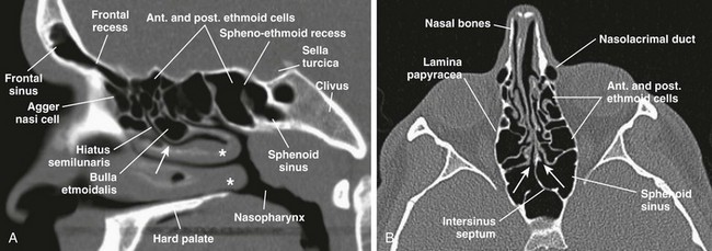
For a ct scan of the sinuses the patient is most commonly positioned lying flat on the back. Sinuses ct brain bone windows the sphenoid sinus and ethmoid air cells are continuous with the nasal airways the mastoid air cells are continuous with the middle ear frontal sinuses ct brain bone windows. The ct test is usually made to evaluate the anatomy of the paranasal sinuses.
The module interface is meant to mimic a radiology workstation with adjacent image scrolling via arrow keys and or mouse wheel button. Welcome to interactive ct sinus anatomy. The patient may also be positioned face down with the chin elevated.
Imaging the paranasal sinuses is routine in clinical practice to evaluate for various sinus pathology non specific facial pain and pre operative planning for functional endoscopic sinus surgery fess including post operative follow up. Straps and pillows may be used to help the patient maintain the correct position and to hold still during the exam. Axial view coronal view sagittal view.
 Paranasal Sinuses Radiology Reference Article
 Sinus Disease Categories South Bay Allergy And Asthma

 Paranasal Sinus Anatomy What The Surgeon Needs To Know
 Sinusitis Cancer Therapy Advisor

 Nasal Cavity Anatomy Physiology And Anomalies On Ct Scan

 Paranasal Sinus Anatomy What The Surgeon Needs To Know
 Ct Anatomy Of Para Nasal Sinuses
 Association Between Endoscopic Radiologic And Patient

 Startradiology

 Brain And Face Ct Interactive Anatomy Atlas

 The Radiology Assistant Paranasal Sinuses Mri
 Sinusitis A Head And Neck Surgeon S Perspective

 Headneckbrainspine
 Paranasal Sinus Anatomy
 5 Assessment Of Maxillary Sinus Anatomy On Ct Scans

 Ct Scan To Delineate The Anatomy Of The Coronary Sinus In A
 Brain And Face Ct Interactive Anatomy Atlas
 Nose And Sinonasal Cavities Radiology Key
Startradiology


Posting Komentar
Posting Komentar