Tarsals five irregularly shaped bones of the midfoot that form the foots arch. Dorsal refers to the top surface of the foot whereas plantar takes its name from the fact that the foot is planted on the ground when it is in contact with a surface.
 Charcot Marie Tooth Disease Orthoinfo Aaos
The end of the leg on which a person normally stands and walks.

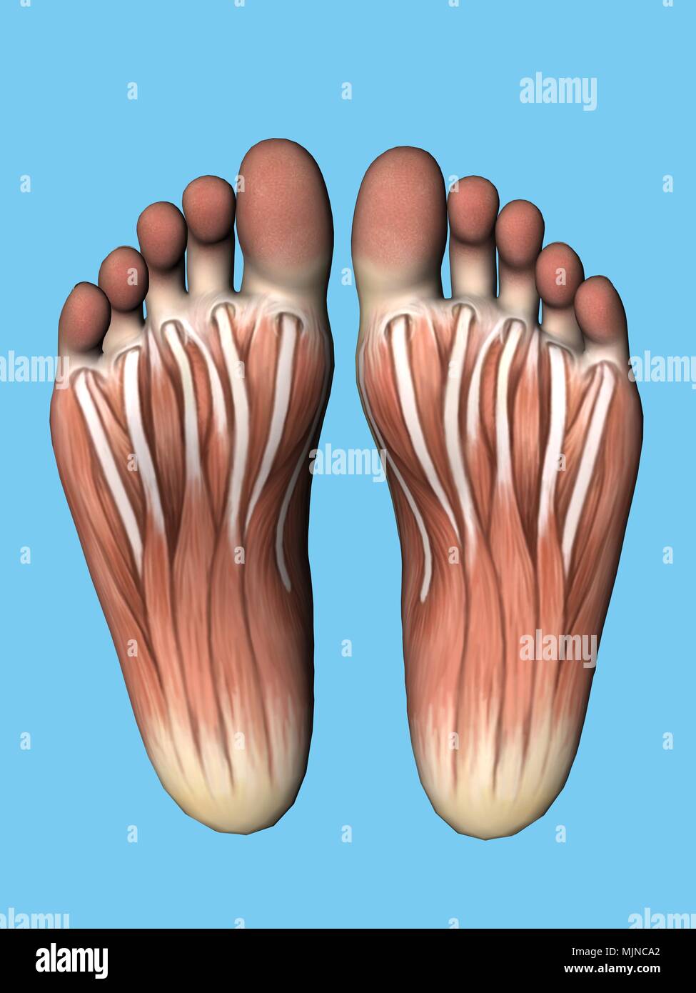
Anatomy of bottom of foot. At the same time the foot must be strong to support more. The midfoot is a pyramid like collection of bones that form the arches of the feet. Therefore plantar refers to the bottom aspect of the foot.
The talus bone supports the leg bones. The anatomy of the foot anatomi cally the foot has two surfaces. The bones of the feet are.
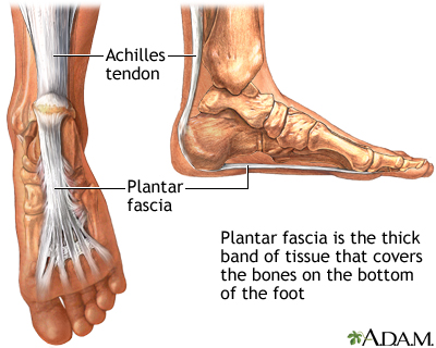
For instance the plantar fascia is an important structure on the bottom of the foot that is important for both the normal function of the foot and for maintaining the normal arch of the foot. The calcaneus which is the bone in your heel. The phalanges which are the bones in your toes.
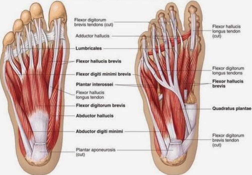
Calcaneus the largest bone of the foot which lies beneath the talus to form the heel bone. Talus the bone on top of the foot that forms a joint with the two bones of the lower leg. The foot is an extremely complex anatomic structure made up of 26 bones and 33 joints that must work together with 19 muscles and 107 ligaments to execute highly precise movements.
The cuneiform bones the navicularis and the cuboid all of which function to give your foot. It is also a common cause of heel pain also known as plantar fasciitis. Below the juncture of these bones are the arches of the foot which are three curves at the bottom of the foot that makes walking easier and less taxing for the body.
The feet are divided into three sections. The talus which is the. Picture of foot anatomy detail.
These arches the medial arch lateral arch and fundamental longitudinal arch are created by the angles of the bones and strengthened by the tendons. The hindfoot forms the heel and ankle. The forefoot contains the five toes phalanges and the five longer bones metatarsals.
Dorsal and plantar see fig. The metatarsals which run through the flat part of your foot.
Anatomy Of The Foot Medical Illustration Medivisuals
 Contemporary Foot Anatomy Bottom Gift Image Of Internal
 Plantar Fasciitis Medlineplus Medical Encyclopedia
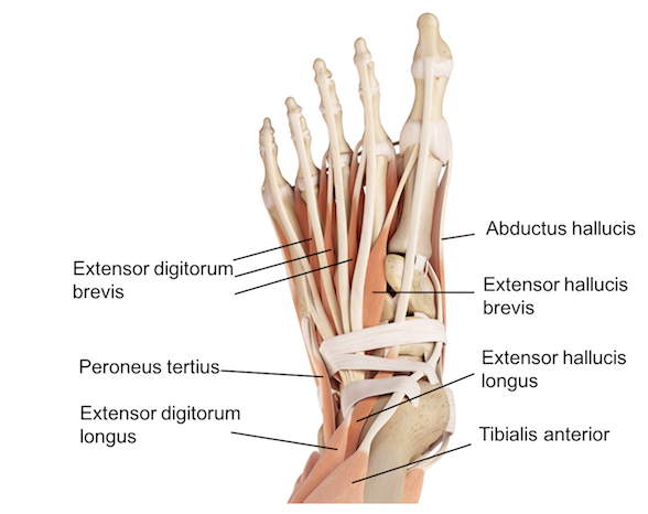
![]() Dorsal Muscles Of The Foot Anatomy And Function Kenhub
Dorsal Muscles Of The Foot Anatomy And Function Kenhub
 Developing Strength Stability In The Foot Ankle And
:max_bytes(150000):strip_icc()/heelpainfinal-01-5c86a48246e0fb00014319ff.png) Heel Pain Causes Treatment And When To See A Doctor
Heel Pain Causes Treatment And When To See A Doctor
 Foot Pain Diagnosis Achilles Tendinitis Causes Home
Anatomy Of The Bottom Of Your Foot Graphjam Funny Graphs
 Athletic Foot Injuries Background Epidemiology Functional
 Nerve Blocks Of The Foot And Ankle Down East Emergency
 Blog Lowry Mcferrin S Website
 Anatomy Of The Foot And Ankle Orthopaedia
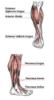
 Muscles Of The Lower Leg And Foot Human Anatomy And
 Cousin Jehan Vintage Bottom Of Foot Vector Anatomy Art Clipart Royalty Free Clipart 403137
![]() Dorsal Muscles Of The Foot Anatomy And Function Kenhub
Dorsal Muscles Of The Foot Anatomy And Function Kenhub
 Massaging Our Sole For Over All Health Personal
 How To Release Your Plantar Fascia Helps Plantar Fasciitis
 Developing Strength Stability In The Foot Ankle And
 Ankle Foot Anatomy
 Sole Of Foot
 Ball Of Foot Pain Do The Bottoms Of Your Feet Toes Hurt
 Anatomy Of The Foot Footmaxx
 Ball Of Foot Pain Relief Management Dr Scholl S
 Nerves Of The Leg And Foot Interactive Anatomy Guide
 Ace Prosource August 2016 Functional Anatomy Series
Plantar Fasciitis And Bone Spurs Orthoinfo Aaos
 Ball Of Foot Pain Do The Bottoms Of Your Feet Toes Hurt
 Ball Of Foot Pain Do The Bottoms Of Your Feet Toes Hurt
 Rheumatoid Arthritis In Feet Symptoms Treatments And More
 Anatomy Bottom View Of Foot Stock Photo 183638170 Alamy
:max_bytes(150000):strip_icc()/footpainfinal-01-d507e82b3e844d068c0089cbb7004d76.png) Foot Pain Causes Treatment And When To See A Doctor
Foot Pain Causes Treatment And When To See A Doctor
 Notes On Anatomy And Physiology Using Imagery To Relax The


Posting Komentar
Posting Komentar