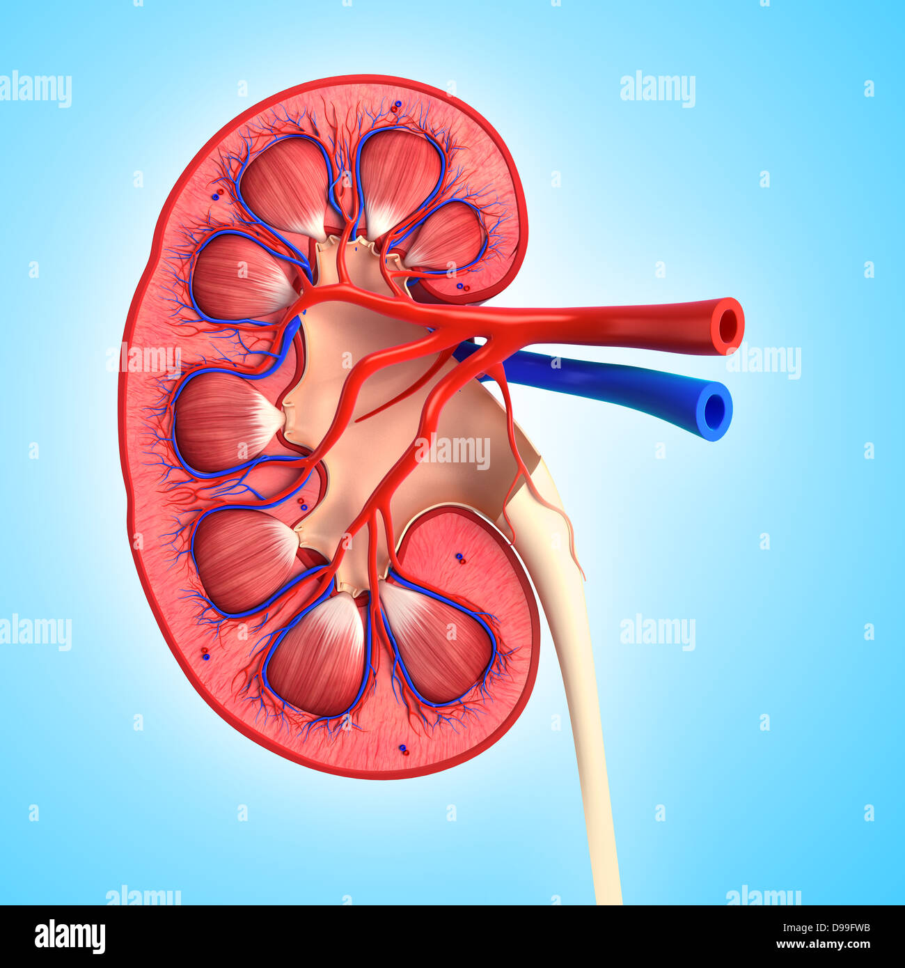
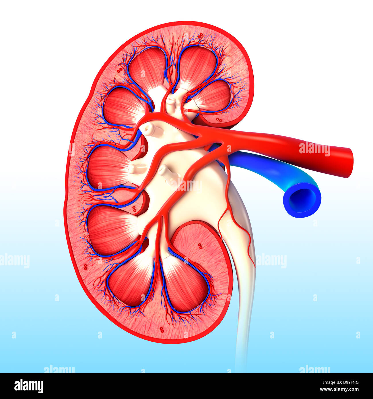
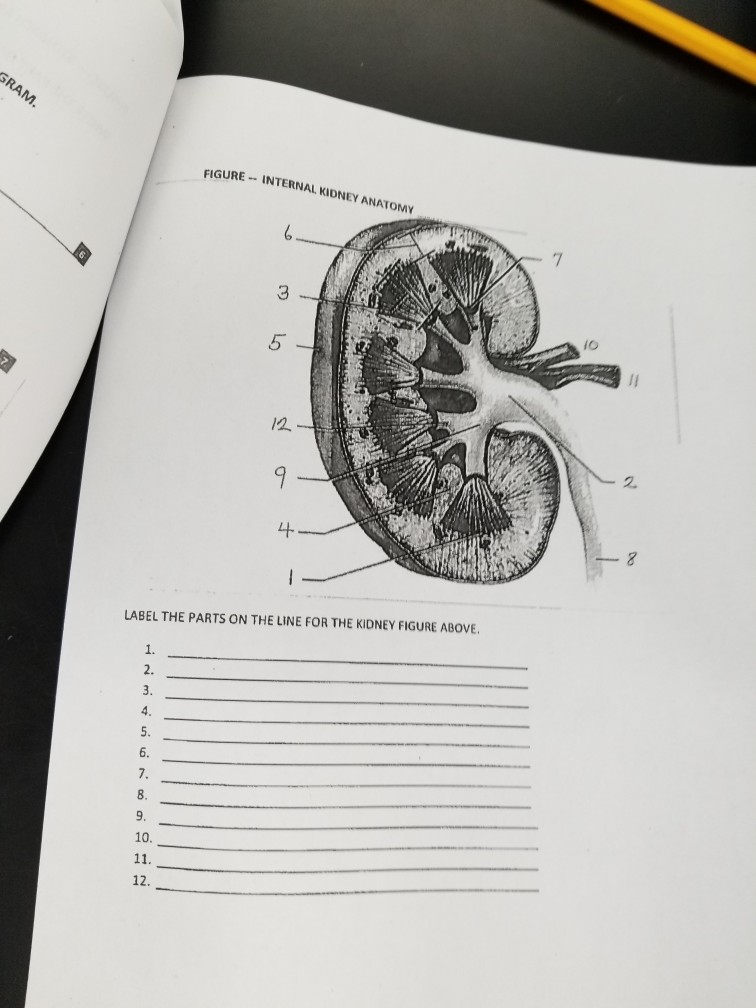
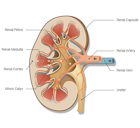
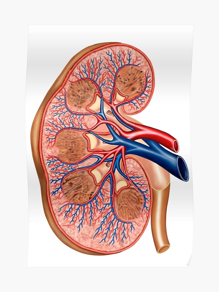
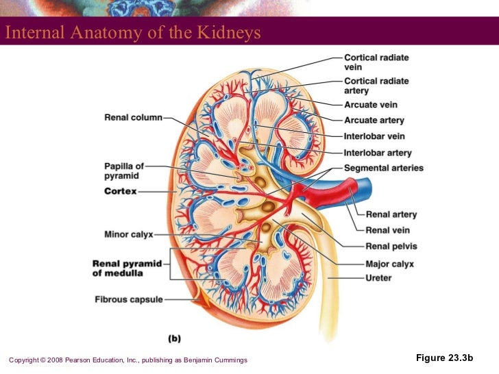
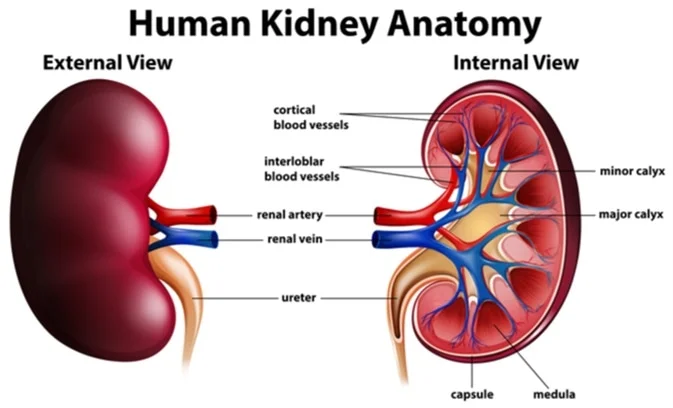
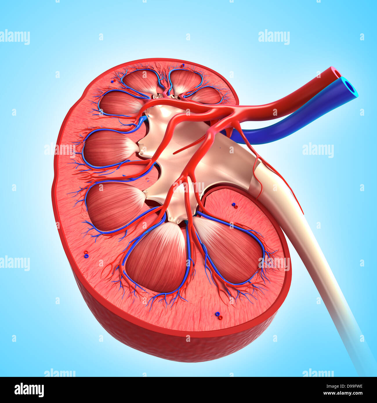
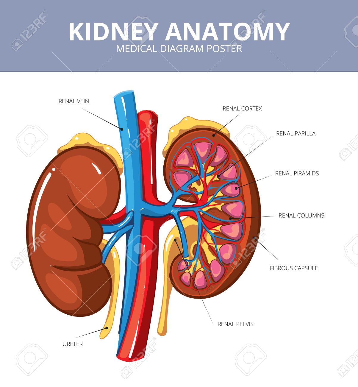
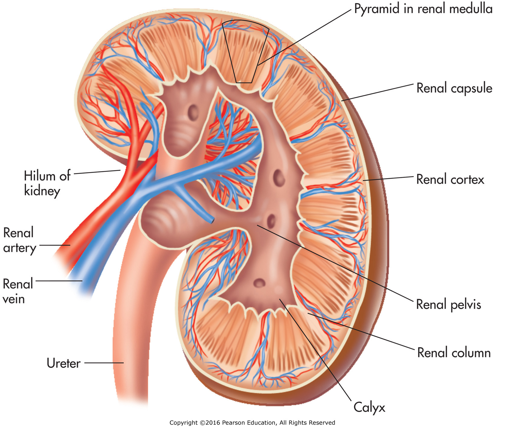
In the medulla 5 8 renal pyramid s are separated by connective tissue renal columns. The kidneys are the main organs of the urinary system and are primarily responsible for removing toxins and other metabolic wastes from the blood.
 Cross Section Of Internal Anatomy Of Kidney Travel Mug
Numerous tubules and ducts make up the pyramids which gives the pyramids a striated appearance especially when viewed microscopically.
Internal kidney anatomy. The renal columns are connective tissue extensions that radiate downward from the cortex through the medulla to separate the most characteristic features of the medulla. Internal anatomy of the kidney overview the main unit of the medulla is the renal pyramid. These curve around the base of the pyramid as the arcuate arteries.
The renal columns are connective tissue extensions that radiate downward from the cortex through the medulla to separate the most characteristic features of the medulla. A frontal section through the kidney reveals an outer region called the renal cortex and an inner region called the medulla figure 2. Internal anatomy a frontal section through the kidney reveals an outer region called the renal cortex and an inner region called the renal medulla figure 2512.
From the arcuate arteries arise a series of branches called the interlobular arteries in the cortex of the kidney. Kidney anatomy encompasses all the internal and external tissue components that collectively form the structure of the kidney. The interlobular veins flow into the arcuate veins the interlobar veins and then the renal vein.
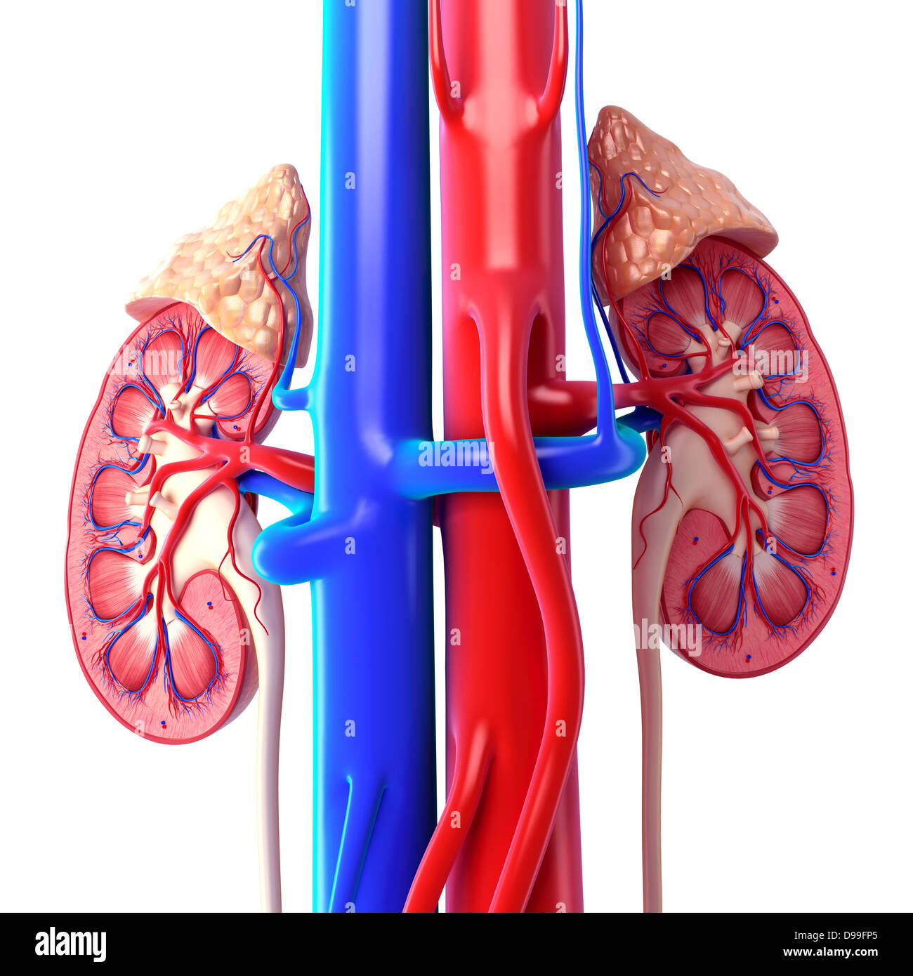
This medial pocket contains the large blood vessels that pass in and out of the kidney and the tubes that conduct urine to the ureters and bladder. Next to the medulla is the renal sinus. There are 8 18 renal pyramids in each kidney that on the coronal section look like triangles lined next to each other with their bases directed toward the cortex and apex to the hilum.
A frontal section through the kidney reveals an outer region called the renal cortex and an inner region called the medulla link. Renal internal anatomy kidney.
Human Body Internal Organs Stomach And Lungs Kidneys And
 Anatomy Of Kidney Internal View In Different Form Stock
 Anatomy Of Kidney Internal View In Different Form Stock
 Internal Anatomy Of Kidney Diagram Quizlet
 Cross Section Of Internal Anatomy Of Kidney Framed Art Print

 Cross Section Of Internal Anatomy Of Kidney Poster
 Renal Artery Wikipedia
 Anatomy Of Kidney Internal View In Different Form
 Kidney Anatomy Detailed Renal Nephron Urinary System
 Anatomy Of Kidney Internal View In Different Form Stock

 Internal Anatomy Of The Kidney Diagram Quizlet
![]() Vector Isolated Illustration Of Kidney Stock Vector
Vector Isolated Illustration Of Kidney Stock Vector
 Human Human Anatomy Urinary System

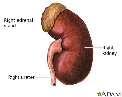
 Anatomy Of The Kidneys Ureter And Bladder Basicmedical Key
 Renal Internal Anatomy Kidney
 Anatomy Of The Kidney
 Anatomy Of Kidney Internal View In Different Form Stock
 Human Kidney Anatomy Kidney Medical Science Vector Illustration
 Kidney And Bladder Urinary System Internal Organs Anatomy Body
 Kidney In Heart Failure Color Atlas And Synopsis Of Heart
 Anatomy Excretory System Science Olympiad Student Center Wiki
 Kidney Medical Vector Diagram Poster Internal Organ Artery
 The Internal And External Anatomy Of The Kidney Biology


Posting Komentar
Posting Komentar