Colorado knee specialist dr. Atlas of knee mri anatomy.
 Knee Mri Part 1 Normal Anatomy For Orthopaedic And Radiology Residents
Anatomy of the knee can be complicated and hard to understand.

Knee mri anatomy. Pain swelling and warmth in any of the bursae of the knee. Bursitis often occurs from overuse or injury. Through the use of magnetic resonance imaging clinicians can diagnose ligament and meniscal injuries along with identifying cartilage defects bone fractures and bruises.
This tool is at the same time useful for the training and teaching of the anatomy. Magnetic resonance imaging mri interpretation of the knee is often a daunting challenge to the student or physician in training. Use the mouse to scroll.
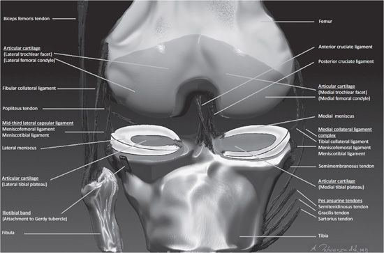
After all an entire year of fellowship training is dedicated to musculoskeletal imaging. This webpage presents the anatomical structures found on knee mri. Each anatomical structure is labelled interactively.
This atlas of cross sectional anatomy of the knee is based on imagery by magnetic resonance mri. Robert laprade discusses how to read an mri of a normal knee. Normal mri anatomy of the knee 639 the fcl biceps femoris bursa is found lateral to the distal fcl and insinuates anterior and antero medial in relation to this ligament.
Collection of fluid in the back of the knee. Click on a link to get t1 coronal view t2 fatsat axial view t2 fatsat coronal view t2 fatsat sagittal view. Anatomy of the knee mri atlas of the human body using cross sectional imaging.
 Knee Mri Radiology Key

 Example Of Mri Slice Mid Thigh With The Knee Extensors And
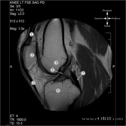
 Mri Knee Anatomy Knee Sagittal Anatomy Free Cross
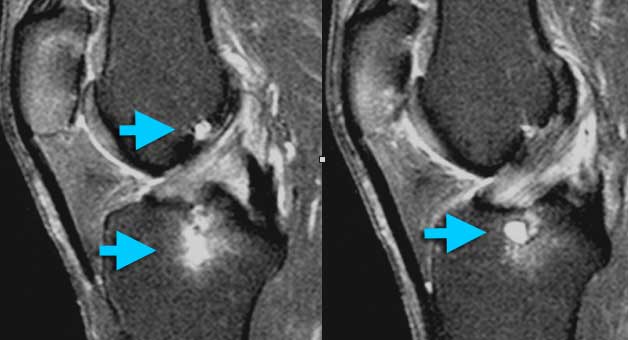 The Radiology Assistant Knee Non Meniscal Pathology

 The Knee Mri Atlas Of Anatomy In Medical Imagery
 Mri Knee Google Search Knee Mri Anterior Cruciate
 Mri Knee Unidad Especializada En Ortopedia Y Traumatologia
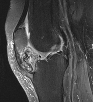 Hoffa S Fat Pad Abnormalities Knee Pain And Magnetic
The Knee Mri Atlas Of Anatomy In Medical Imagery


 Radiology Images
 Knee Anatomy Mri Knee Coronal Anatomy Free Cross
 The Knee Mri Atlas Of Anatomy In Medical Imagery
Stanford Msk Mri Atlas C 2019
Magnetic Resonance Imaging Of The Knee And Correlation With
 Ppt Posterolateral Corner Of The Knee Mri Anatomy
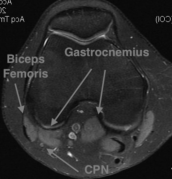
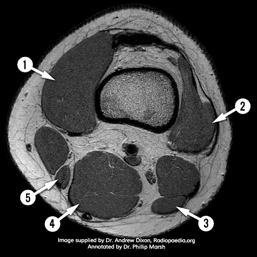 Mri Knee Axial Anatomy Quiz Radiology Case
 Department Of Anatomy Med Univ Of Warsaw Poland Knee
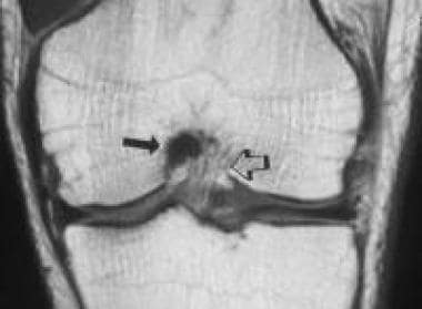 Mri For Posterior Cruciate Ligament Injuries Overview
 Acl Anatomy Eorif
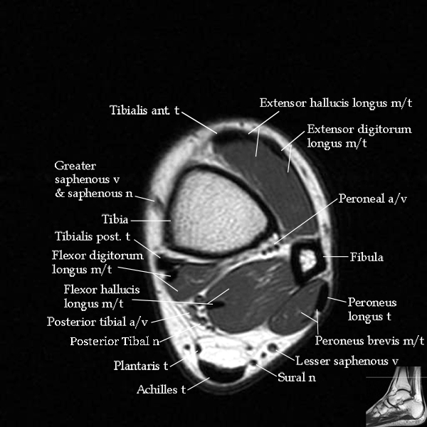 Mri Ankle Anatomy


Posting Komentar
Posting Komentar