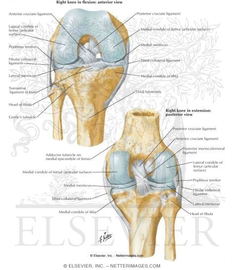
Annular ligament from. The lateral radial collateral ligament lclrcl complex is a major lateral stabilizer of the elbow joint and resists varus stress.

Collateral analytics ca develops real estate analytic products and tools to support financial institutions institutional and retail investors as well as property capital market activities.
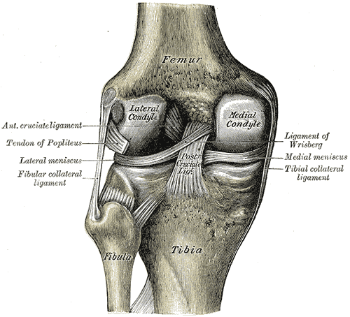
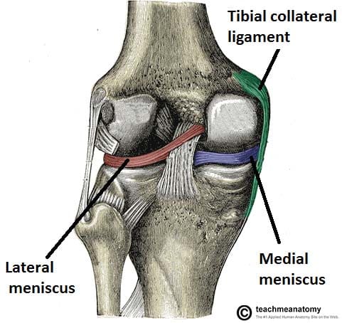
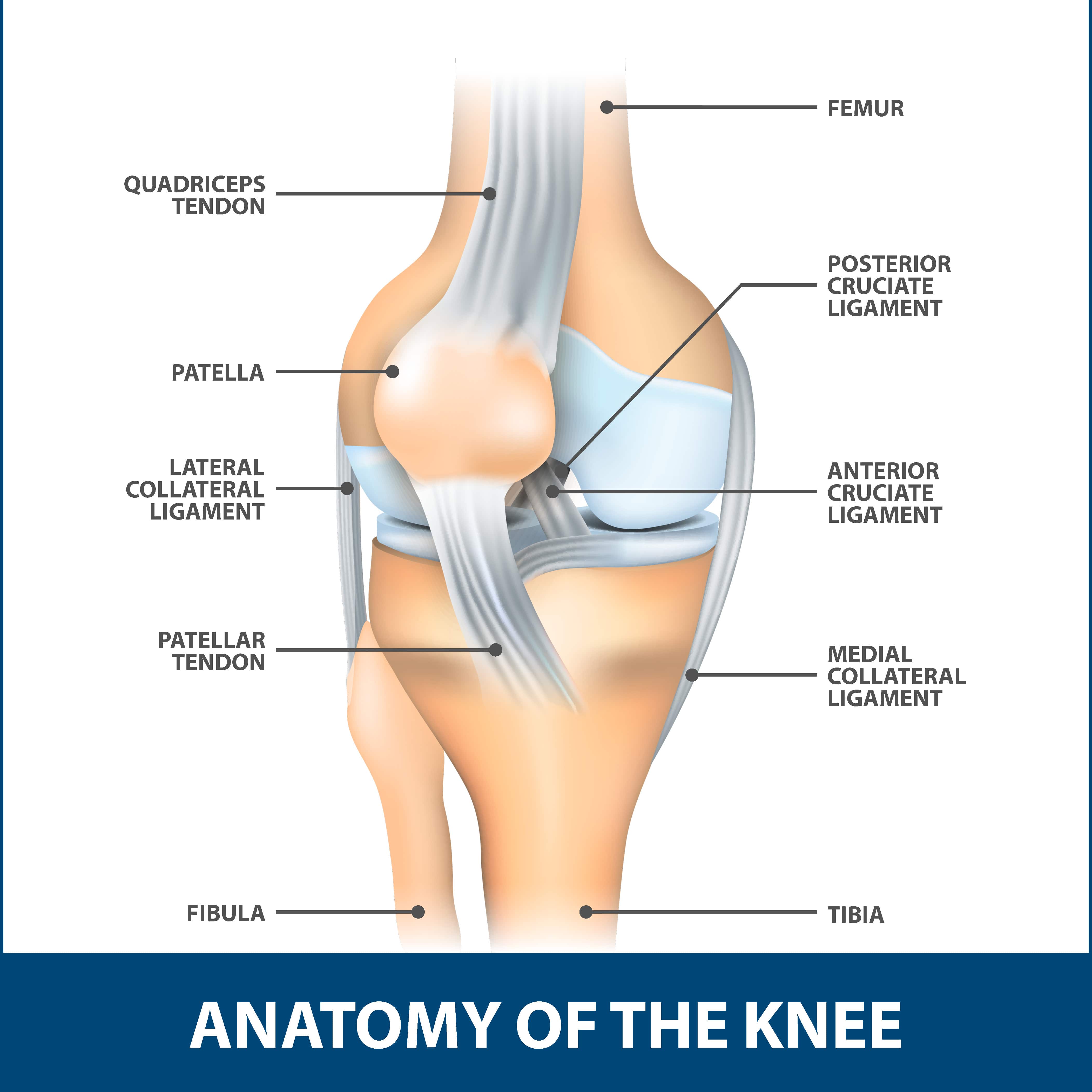
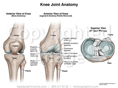
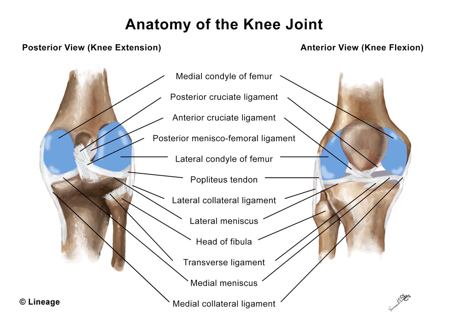
Collateral anatomy. In anatomy a collateral is a subordinate or accessory part. The structures that are considered static stabilizers of the medial knee are the superficial mcl the deep mcl and the posterior oblique ligament. The medial collateral ligament is recognised as being a primary static stabiliser of the knee and assists in passively stabilising the joint.
Gross anatomy the lcl is a y shaped ligamentous complex composed of three parts 1 2. The axon collateral can be a part of feedback mechanism which creates a connection with nearby inhibitory neurons and thus they can be involved in regulation of the neuron over excitation. These are often contact injuries but not always.
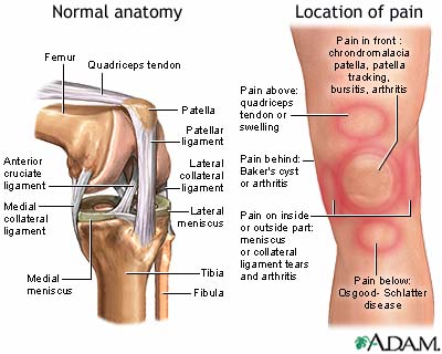
The superficial mcl is the primary static medial stabilizer of the knee situated in the second layer according to warren and marshalls three layer concept. Injuries to the collateral ligaments are usually caused by a force that pushes the knee sideways. A collateral is also a side branch as of a blood vessel or nerve.
The dendrites accept the signal received from the other nerve cells and the axon carried signals to the axon terminals. The mcl also prevents an anterior movement of the tibia and hyperextension. However several of their branches can become important collateral pathways if occlusion occurs in the internal carotid or vertebral arteries.
A collateral is also a side branch as of a blood vessel or nerve. The collateral ligaments medial mcl and lateral lcl are found on the sides of your knee. Two c shaped pieces of cartilage called the medial and lateral menisci act as shock absorbers between the.
Learn about financial products learn about real estate products. Indirect subsidiary or accessory to the main thing. The branches of the external carotid artery are the ascending pharyngeal the superior thyroid the lingual the external maxillary the occipital the facial the posterior auricular.
The medial and lateral collateral ligaments prevent the femur from sliding side to side. A side branch of a nerve axon or blood vessel. When stress is applied this ligament aids control in transferring the joint through a normal range of movement.
The axon terminals send signals to the next nerve cell and so forth.
 Fibular Collateral Ligament Wikipedia
 Image Result For Medial Collateral Ligament Elbow Elbow
 Figure 1 From Arthroscopically Accessible Anatomy Of The
 Elbow Anatomy Biomechanics Shoulder Elbow Orthobullets
 Mri Musculo Skeletal Section Radial Collateral Ligament
 14190 01b Abdominal Vasculature With Collateral Flow

 Understanding The Medial Ulnar Collateral Ligament Of The
 Collateral Ligament Cl Injury Aftercare Medlineplus
 The Knee Joint Articulations Movements Injuries
 End Arteries Anastomosis And Collateral Circulation
 Collateral Anatomy Exhibits
 Cruciate And Collateral Ligaments Of Right Knee Joint Knee
 Joseph D Foley Iii Md On Twitter Great Talk Earlier
 Thumb Approach Dorsoulnar To Mcp Joint Of Thumb Ao
 Lateral Collateral Ligament Florida Orthopaedic Institute
 Knee Joint Anatomy

 Knee Human Anatomy Function Parts Conditions Treatments
 Understanding The Medial Ulnar Collateral Ligament Of The
 Upper Limb Vascular Anatomy Collateral Arterial Blood
 Collateral Ligament Tear Orthopedics Medbullets Step 2 3
 Collateral Ligaments Of The Knee Joint Patellar Tendon
 Ulnar Collateral Ligament Anatomy Park Sports Physical Therapy
 Normal Anatomy Of Left Knee Collateral Ligament Medical

 Anatomy Of The Elbow Elbow Anatomy


Posting Komentar
Posting Komentar