The lateral radiograph side aspect of the lumbar spine shows the vertebral bodies and intersomatic spaces the intervertebral faces of the vertebral bodies and laminae. Radiographic anatomy of the skeleton.
 Pediagenosis
The radiographic anatomy and patient positioning.

Radiograph anatomy. Medical students typically spend some time studying radiographic anatomy during their general educations and certain medical specialists may go on to study it extensively such as radiographers orthopedic surgeons and dentists. Radioanatomy x ray anatomy is anatomy discipline which involves the study of anatomy through the use of radiographic films. Check for errors and try again.
Radiographic anatomy fractures and luxations involving the temporomandibular joint. Unable to process the form. The online sira module consisted of text and visual aids introducing the identification of normal radiographic anatomy on intraoral and extraoral radiographic images.
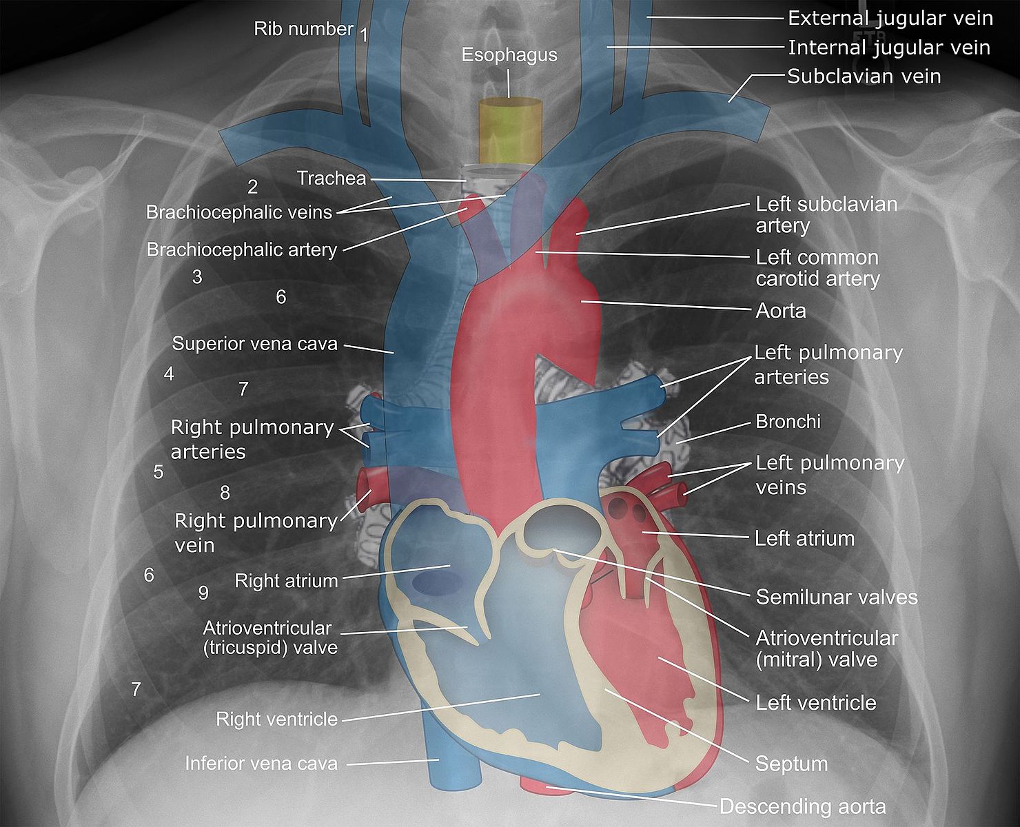
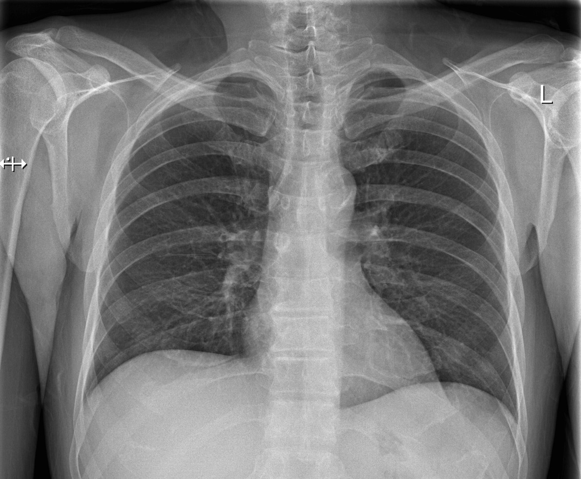
Chest x ray anatomy many structures of the chest are readily visible on a chest x ray but others are difficult to see. We would like to show you a description here but the site wont allow us. Other anatomical structures such as the pleura only become clearly visible when abnormal.
The x ray film represents two dimensional image of a three dimensional object due to the summary projection of different anatomical structures onto a planar surface. In fact some important structures such as the phrenic nerve are not visible at all. Radiographic anatomy is a branch within the discipline of anatomy which involves the study of anatomy through the use of radiographic films also known as x rays.
The three quarters radiograph oblique lumbar spine aspect is particularly useful for identifying the zygapophysial. The radiographic anatomy of owls is similar to other birds. Sarah nemanic dvm phd ms dacvr.
The results of the pre test demonstrated that faculty had knowledge of radiographic anatomy 45 90 with a median score of 65.
Anatomy Of An Hand Xray Purposegames
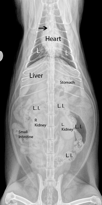 Learn How To Interpret Cat Radiographs X Rays Long
 Lateral Chest Radiograph Anatomy Normal Lateral Chest X
 Radiographic Anatomy Of Adult Hand Orthopaedicsone
 Mandible X Rays
 Cervical Spine Radiographic Anatomy Radiologypics Com
 Normal Radiographic Anatomy Of The Knee Radiology Case
 Plain Film X Ray Principles Interpretation Teachmeanatomy
 Radiographic Anatomy An Overview Sciencedirect Topics
 Startradiology
 Radiology Basics Abdomen Anatomy
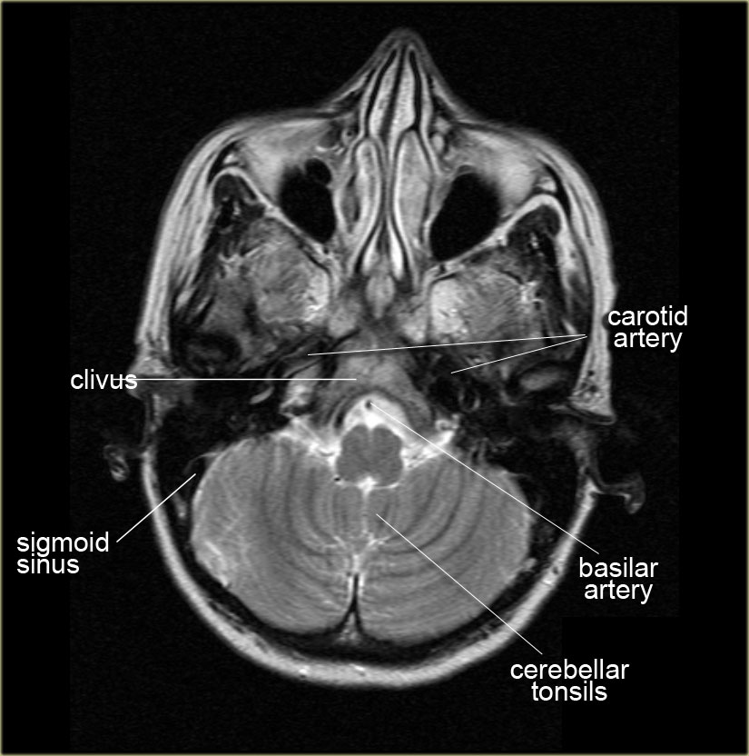 The Radiology Assistant Brain Anatomy
 Carpus Equine Anatomy Radiology Small Animal Hospital
 Anatomy On The Pa And Lateral Radiograph The Medical Media
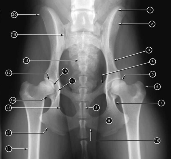
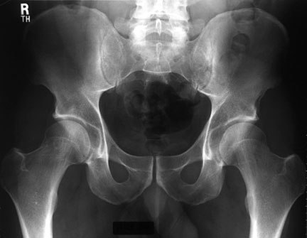
 Xray Anatomy Of The Hip Review Xray Anatomy Of The Hip
 Radiographic Anatomy Of The Appendicular Skeleton Veterian Key
 Radiograph Anatomy X Ray Stock Photos And Images Age Fotostock
 Labeled Cervical Spine Xray Anatomy Odontoid View Anatomy
 Foot X Rays
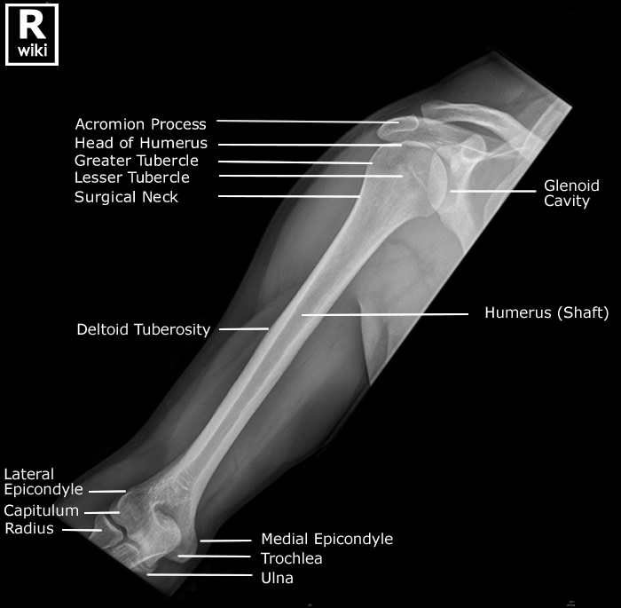 Radiographic Anatomy Of The Humerus Radiologypics Com
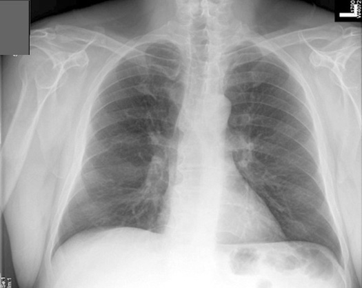
 Practice Chest X Ray Interpretation
 Hand X Ray Pa Hand Radtechonduty
 Radiographic Anatomy Of The Skeleton Table Of Contents
 Radiology Anatomy
 Thorax Radiologic Anatomy
 Normal Radiographic Anatomical Landmarks


Posting Komentar
Posting Komentar