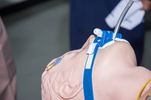
Endotracheal intubation is a basic skill that every first responder must master. A keen understanding of airway anatomy can make the process of intubating a patient much easier.
 Pin On Rt
Nasotracheal intubation is an alternative approach to orotracheal intubation.
Anatomy intubation. And while it does pose some risks it is also safe with the right technique and diligent attention to the patient. The larynx is a cartilaginous structure slung from the hyoid bone by the hyothyroid membrane. The lower airway consists of the subglottic larynx the trachea and the bronchi.
The most widely used route is orotracheal in which an en. Navigation best viewed on larger screens. The nasal fossa is bounded laterally by inferior middle and superior turbinate bones.
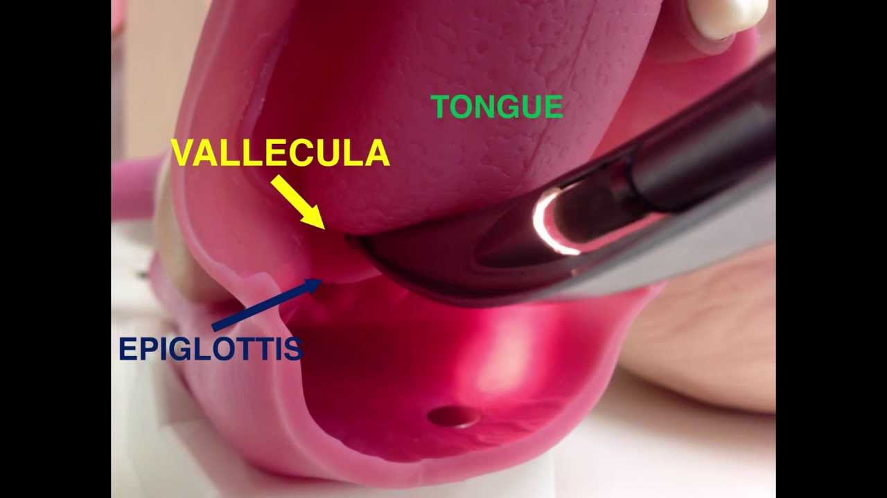
The presentation begins with anthony lewis outlining basic upper airway anatomy in particular the anatomy of the tongue and its attachments and the cricoid membrane. It is frequently performed in critically injured ill or anesthetized patients to facilitate ventilation of the lungs including mechanical ventilation and to prevent the possibility of asphyxiation or airway obstruction. Intro to intubation 01 sequen.
The nasal fossae are divided by the midline cartilaginous septum and medial portions of the lateral cartilages fig. Try using search on phones and tablets. The two nasal fossae extend from the nostrils to the nasopharynx.
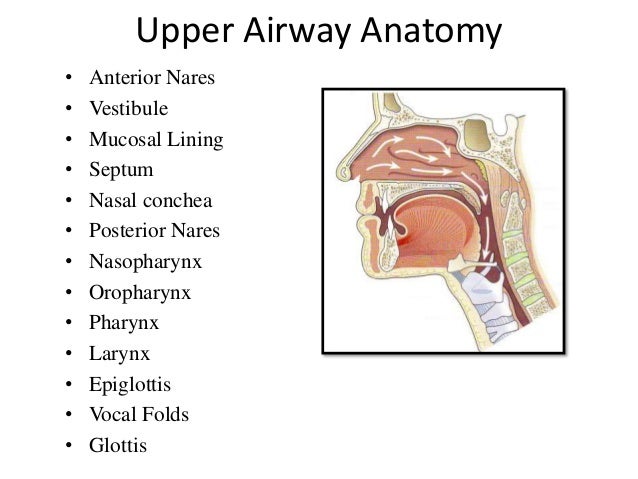
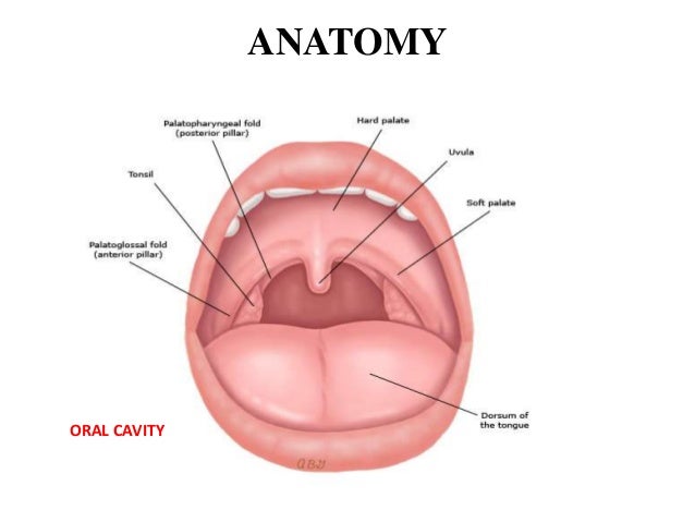
The ligaments of the larynx antero lateral view. Intubation anatomy the upper airway comprises the nasal and oral cavities the pharynx and the larynx. He explains how these structures can affect the airway and the principles behind basic airway manoeuvres.
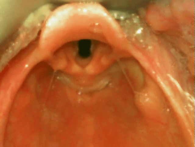
The larynx is the key anatomical structure that needs to be identified when carrying out intubation. It comprises of numerous separate cartilages held together with connective tissue. Tracheal intubation usually simply referred to as intubation is the placement of a flexible plastic tube into the trachea to maintain an open airway or to serve as a conduit through which to administer certain drugs.
 Ligaments And Membranes Of The Larynx Google Search
 Tracheal Intubation Wikipedia
 Functional Anatomy And Physiology Of Airway Intechopen
Endotracheal Intubation With Laryngoscope Medical Chart
 Airway Intubation Stock Photos Airway Intubation Stock
 Paediatric Airway Anatomy Paediatric Emergencies
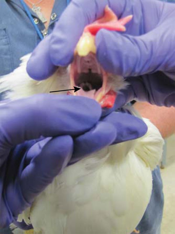 Endotracheal Intubation And Oral Gavage In The Domestic
 Endotracheal Intubation In Oral Maxillofacial Surgery
 Week 1 2 Generallyvolatile
 Pin On The Greater Mind
Airway Anatomy And Endotracheal Intubation The Basics

/intubation-021-5a299722e258f8003693b043.png) What Is Intubation And Why Is It Done
What Is Intubation And Why Is It Done
 Infant Newborn Baby Endotracheal Intubation Anatomy Training
 Intubation Preparation And Equipment Paediatric Emergencies

 Airway Devices 01 Direct Laryngoscopy
 Functional Anatomy And Physiology Of Airway Intechopen
 Functional Anatomy And Physiology Of Airway Intechopen
 Functional Anatomy And Physiology Of Airway Intechopen
 Intubation And Upper Airway Management Thoracic Key
 Endotracheal Intubation Review Of Critical Care Medicine
 Intubation And Mechanical Ventilation 22 Dr Virbhan Balai
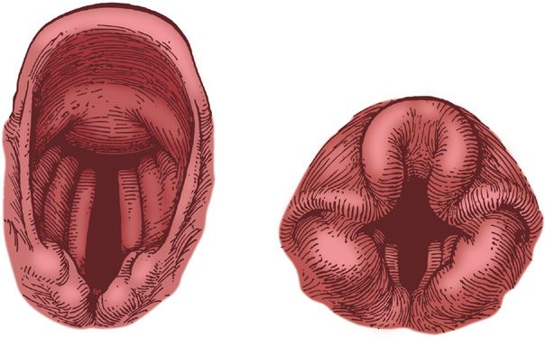
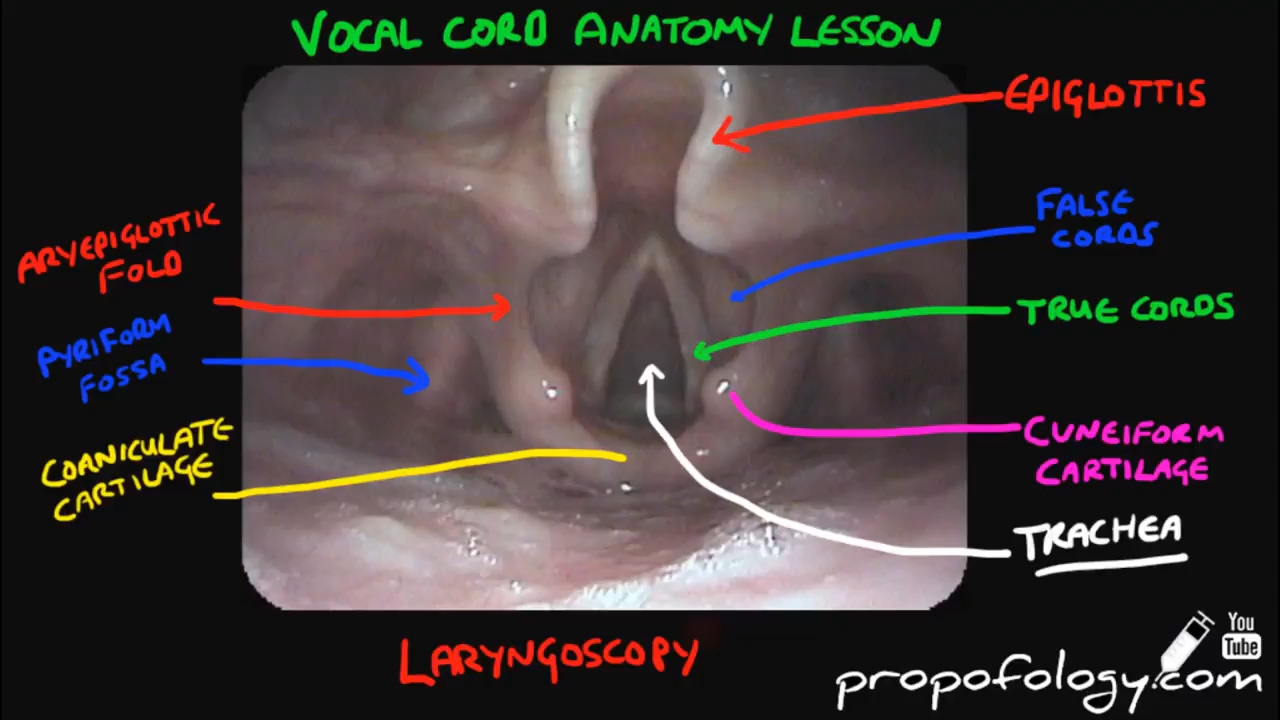 Vocal Cord Anatomy In 2 Minutes Anatomy
 Chapter 38 Airway Management Principles And Practice Of
 Chapter 6 Essential Anatomy Of The Airway Emergency
 Pin On Clinical Skills

 Regional And Topical Anesthesia For Awake Endotracheal
 Improper Endotracheal Intubation Doctor Stock


Posting Komentar
Posting Komentar