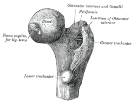
The head forms a ball and socket joint with the hip at the acetabulum being held in place by a ligament ligamentum teres femoris within the socket and by strong surrounding ligaments. Lesser trochanter smaller than the greater trochanter.
 Anatomy Of The Femur Bone The Pillar Of Support For The
The head of the femur articulates with the acetabulum in the pelvic bone forming the hip joint while the distal part of the femur articulates with the tibia and kneecap forming.

Anatomy of the femur bone. In humans the neck of the femur connects the shaft and head at a 125 angle. Start studying bone anatomy of the femur. The area of the bone supports the strongest muscle tissue in the body including the hamstrings quadriceps and thigh musculature.
It is commonly known as the thigh bone femur is latin for thigh and reaches from the hip to the knee. It is both the longest and the strongest bone in the human body extending from the hip to the knee. The shaft of the femur is somewhat curved and has a protruding ridge called the linea aspera rough line.
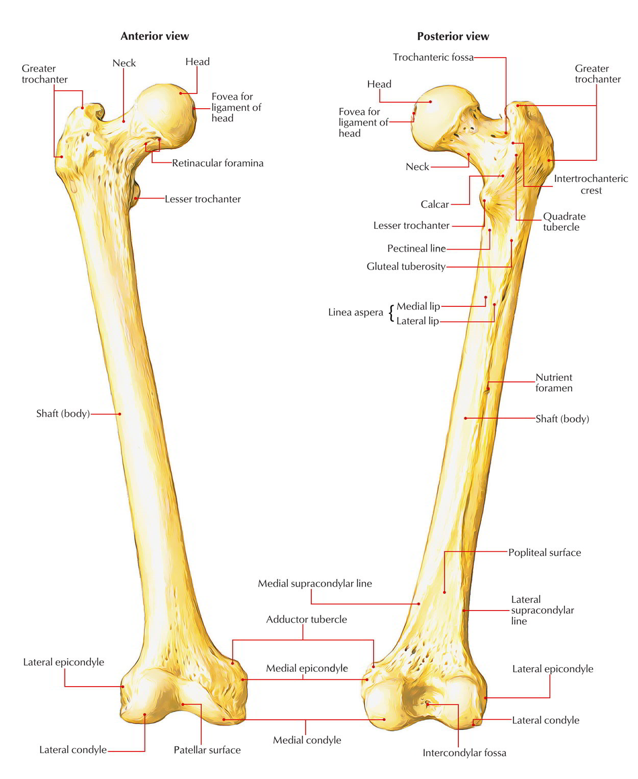
Important features of this bone include the head medial and lateral condyles patellar surface medial and lateral epicondyles and greater and lesser trochanters. Neck connects the head of the femur with the shaft. The femur is the only bone located within the human thigh.
The femur ˈfiːmər pl. Femur also called thighbone upper bone of the leg or hind leg. Femurs or femora ˈfɛmərə or thigh bone is the proximal bone of the hindlimb in tetrapod vertebrates.
Learn vocabulary terms and more with flashcards games and other study tools. The femur is the largest bone in the human body. Greater trochanter the most lateral palpable projection of bone that originates from.
Proximal head articulates with the acetabulum of the pelvis to form the hip joint. The vastus laterallis outer quadricep and adductor magnus inner thigh muscle. Femur or thigh bone is the longest the strongest bone of the body.
A human male adult femur is about 19 inches long and weighs a little more than 10 ounces.
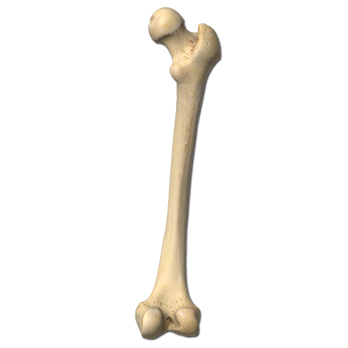
 3d Illustration Of Skeleton Femur Bone Anatomy Stock
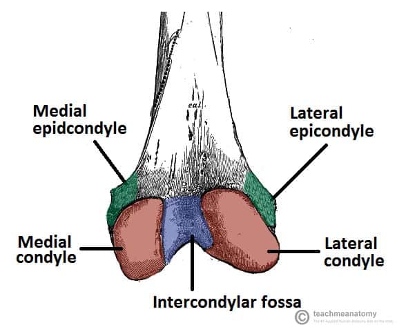 The Femur Proximal Distal Shaft Teachmeanatomy

 Easy Notes On Femur Learn In Just 4 Minutes Earth S Lab
 Femur Bone Honors Anatomy Diagram Quizlet
 The Lower Limbs Human Anatomy And Physiology Lab Bsb 141
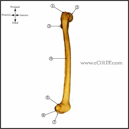 Femur Anatomy Eorif
 Anatomy Of The Horse Osteology
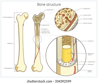
 Femur Bone Structure Stock Vector Illustration Of Health
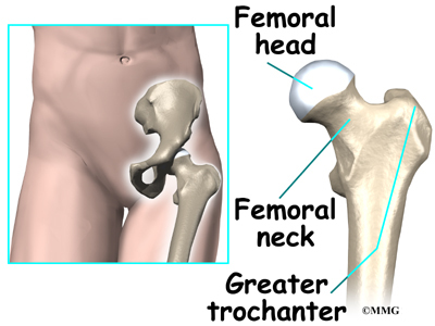
 Anatomy Of The Hip Central Coast Orthopedic Medical Group
 Hip Anatomy Recon Orthobullets
Knee Joint Picture Image On Medicinenet Com
 3d Illustration Of Skeleton Femur Bone Anatomy Stock
 Anatomy Of The Human Proximal Femur Upper Third Of The
 Femur Wikipedia
 Royalty Free Femur Stock Images Photos Vectors Shutterstock
 Femur Labeling Quiz
 Human Femur Anatomy With Porosity And Stiffness At Different
 Femur Wikipedia
 Femur Wikiwand
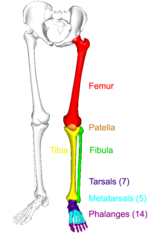
 Bones Of The Leg And Foot Interactive Anatomy Guide

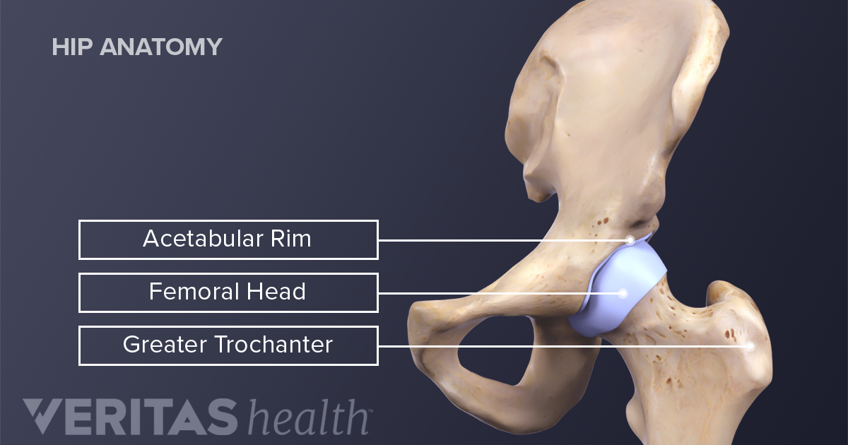 Hip Anatomy


Posting Komentar
Posting Komentar