Welcome to navigating the skull base. Basic anatomy review the bones sutures and fissures that comprise the skull base.
 Skull Base Anatomy By Dr Aditya Tiwari
Ct demonstrates the bony anatomy best while mri has superior soft tissue resolution.

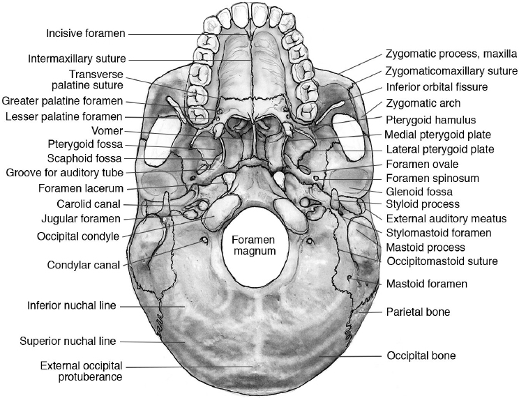
Skull base anatomy. This webpage presents the anatomical structures found on skull base ct. The bones of the skull can be divided into two groups. 5 internal occipital protuberance.
Navigating the skull base. The skull base anatomy is complex and is not directly accessible for clinical evaluation. Surgical exploration without accurate knowledge of anatomy can be catastrophic.
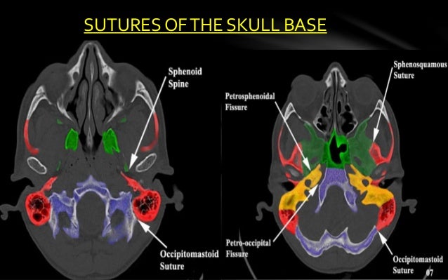
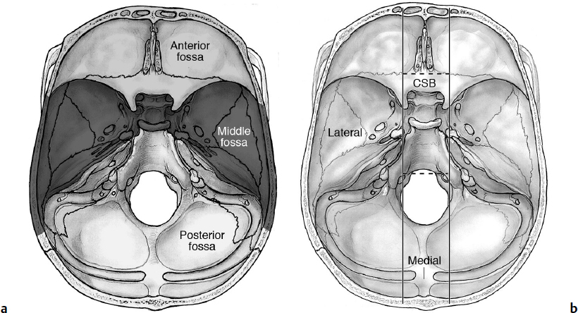
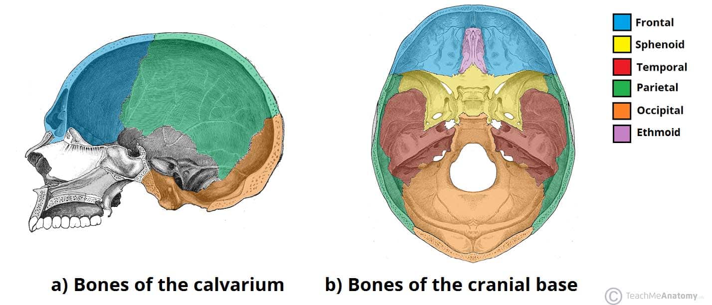
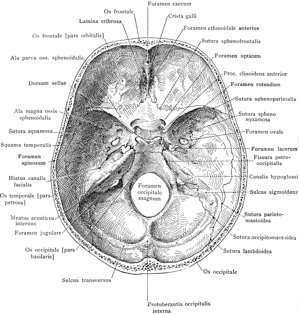
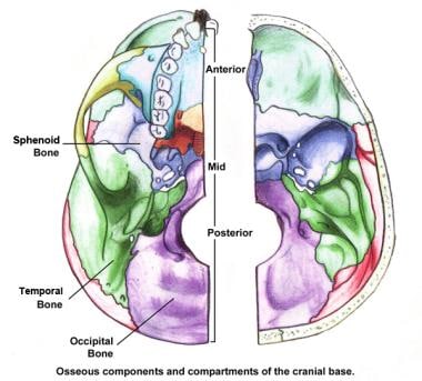
The skull base is made of the paired frontal and temporal bones as well as the ethmoid and occipital bones and these bones form the floors of the anterior middle and posterior cranial fossa. The skull base forms the floor of the cranial cavity that separates brain from facial structures and suprahyoid neck. Navigating the skull base identify the petro occipital fissure to navigate the major structures of the skull base.
To begin click on introduction. 2 superior orbital fissure. Ct anatomy of skull base.
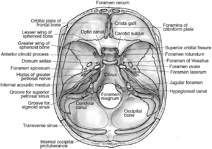
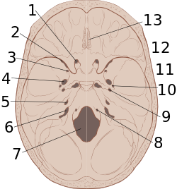
3 anterior clinoid process. The 5 bones that make up the skull base are the ethmoid sphenoid occipital paired frontal and paired temporal bones. 2 superior orbital fissure.
Base of the skull gross anatomy. It separates brain from facial structures and suprahyoid neck. Those of the cranium which can be subdivided the skullcap known as the calvarium and the cranial base and those of the face.
Click the next link at the bottom of each page to step through each section. 3 anterior clinoid process. The cranium the cranium also known as the neurocranium is formed by the superior aspect of the skull.
You can however go directly to any section using the links below. An interactive program for learning skull base anatomy. The base of the skull is perforated by numerous foramina which allow vessels.
Detailed anatomy enter this module for a more detailed review of skull base anatomy. Additionally a poorly defined region termed the central base of skull is often used clinically. There are five bones that make up the base of the skull.
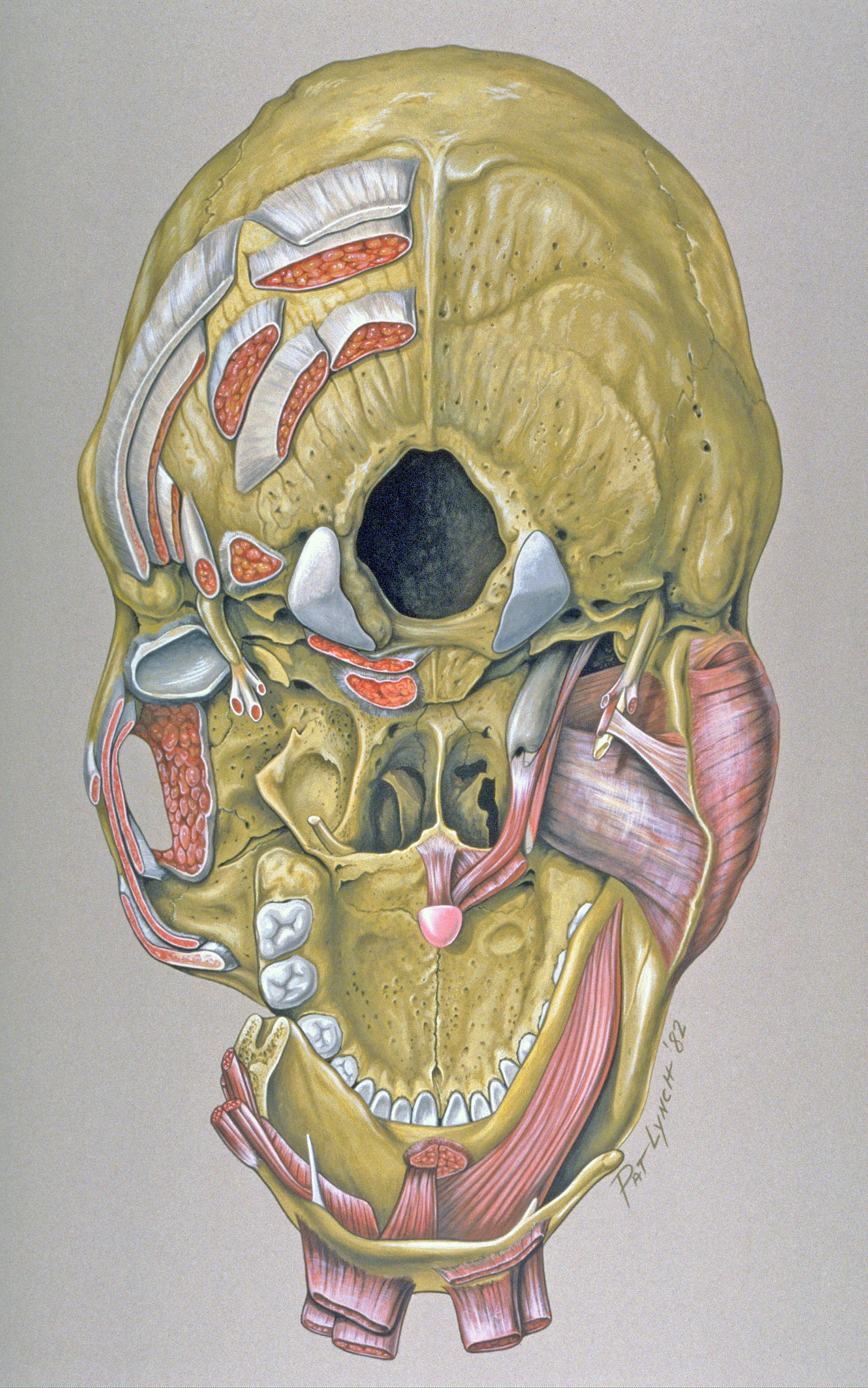
Anatomical knowledge of this particular region is important for under standing several pathologic conditions as well as for planning surgical procedures. 4 mastoid air cells. These bones are separated from each other by sutures.
Working knowledge of the normal and variant anatomy of the skull base is essential for effective surgical treatment of disease in this area. The skull base represents a central and complex bone structure of the skull that forms the floor of the cranial cavity on which the brain lies. Ct anatomy of skull base.
 Anatomy Of The Skull Base And Related Structures Elements
 Skull Base Imaging Anatomy Pathology And Protocols

 Anatomy Of The Skull Base And Related Structures Elements
 Skull Base Anatomy Overview Anterior Skull Base Middle
 Bony Anatomy Of The Orbit And Skull Base
 Anatomy Quiz Skull Base Radiology Case Radiopaedia Org
 Skull Base Osuccc James
 Rhinology Diseases Of The Nose Sinuses And Skull Base
 Anatomy Of The Skull Base And Related Structures Elements
 Skull Base Review And Pathology Veomed Cranial Anatomy
 Skull Anatomical Illustrations
 Base Of The Skull Medatrio
 Anatomy Atlas Part 5 Skull Base
 A 3d Stereotactic Atlas Of The Adult Human Skull Base
 Neurosurgery Transnasal Endoscopic Skull Base And Brain

 Skull Base Anatomy By Dr Aditya Tiwari
 Base Of Skull Wikipedia
 Bones Of The Skull Structure Fractures Teachmeanatomy
 Base Of Skull From Within Clipart Etc
 File Skull Base Anatomy Jpg Wikimedia Commons
![]() Inferior View Of The Base Of The Skull Anatomy Kenhub
Inferior View Of The Base Of The Skull Anatomy Kenhub
 Headneckbrainspine
 Skull Base Imaging Anatomy Pathology And Protocols
Skull Base Anatomical Landmarks Medicalchemy Anatomy
 Skull Base Anatomy Overview Anterior Skull Base Middle

 Surgical Anatomy And Physiology For The Skull Base Surgeon
 Base Of Skull From Above Skull Base Anatomy Physiology
 Imaging Of Paranasal Sinuses And Anterior Skull Base And


Posting Komentar
Posting Komentar