The ossification or formation of the bone starts from three centers. Muscles attachments of the tibia.
It is largely responsible for muscle movement and in distributing and supporting the body weight of the person equally.
Tibial anatomy. Muscles insertions on the tibia tensor fascia latae muscle at gerdys tubercle via the iliotibial band. Quadriceps femoris at the tibial tuberosity. As in other vertebrates the tibia is one of two bones in the lower leg the other being the fibula and is a component of the knee and ankle joints.
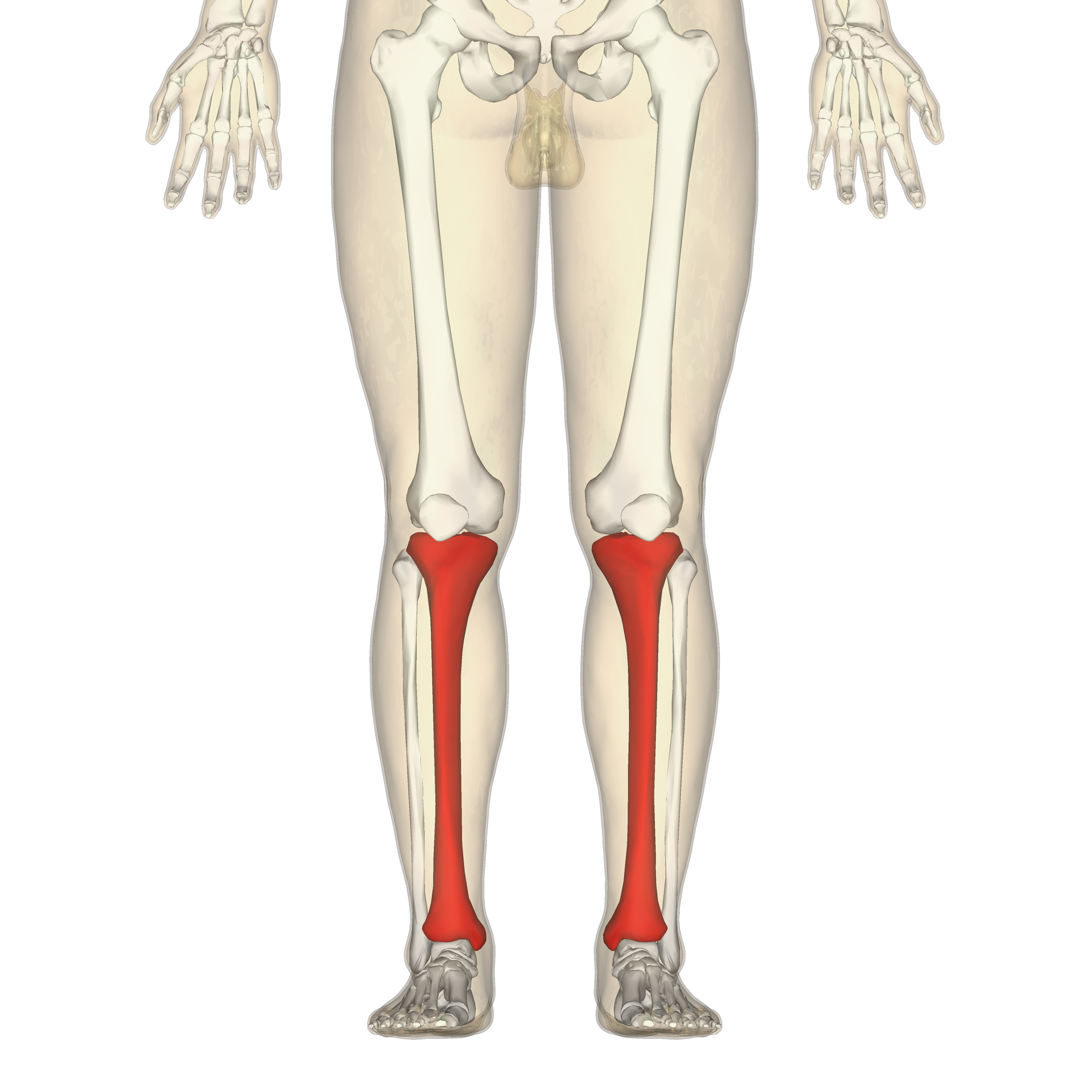
The tibia is the main bone of the lower leg forming what is more commonly known as the shin. Blood supply of the tibia. The tibia is the second largest bone in the body and it is a key weight bearing structure.
Long bones contain bone marrow in a cavity running the length of the shaft. Horizontal head of semimembranosus inserts at medial condyle. The tibia or the shinbone is the second largest bone in the body after the femur bone.
The fibula is smaller and thinner than the tibia. They are divided into a deep and superficial compartment. Sartorius gracilis semitendinosus form a wide insertional slip called.
These two bones connect the ankle to. One in the shaft and one in each extremity. It is situated on the lateral side of the tibia.
Deep popliteus laterally rotates the femur on the tibia to unlock the knee. The tibialis anterior overlaps the anterior tibial vessels and deep peroneal nerve in the upper part of the leg. Articulating at the knee and ankle joints respectively.
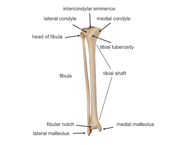
Bony landmarks muscle attachment parts of the tibia. The tibial tuberosity and anterior crest are clearly identifiable landmarks of the shin as they can be easily palpated through the skin. The tibia is also known as the shinbone and is the second largest bone in the body.
The tibia is blood supplied. The tibial nerve innervates all the muscles in the posterior compartment of the leg. Long bones are found on the upper and lower limbs fingers and toes.
In human anatomy the tibia is the second largest bone next to the femur. This muscle is mostly located near the shin. Flexor hallucis longus flexes the big toe and plantar flexes the ankle.
At the proximal end the tibia is widened by the medial and lateral condyles. It expands at its proximal and distal ends. The tibia is a long bone which means it is a limb bone that is longer than it is wide.
Tensor fasciae latae muscle insert into the gerdys tubercle. Approaching the ankle joint the tibia widens slightly in both the medial lateral and anterior posterior planes. There are two bones in the shin area.
It is thick and fleshy above tendinous below. The tibia and fibula or calf bone. On the medial side the tibia forms a rounded bony prominence known as the medial malleolus.
The tibia is a large bone located in the lower front portion of the leg. The tibialis anterior is a muscle in humans that originates in the upper two thirds of the lateral surface of the tibia and inserts into the medial cuneiform and first metatarsal bones of the foot. It acts to dorsiflex and invert the foot.
 The Knee Joint And Leg Yogabody Anatomy Kinesiology And
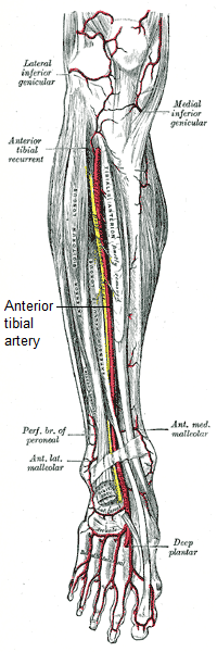
 Anterior Tibial Dorsalis Pedis Arteries Branches Anatomy Tutorial
 Tibia Radiology Reference Article Radiopaedia Org
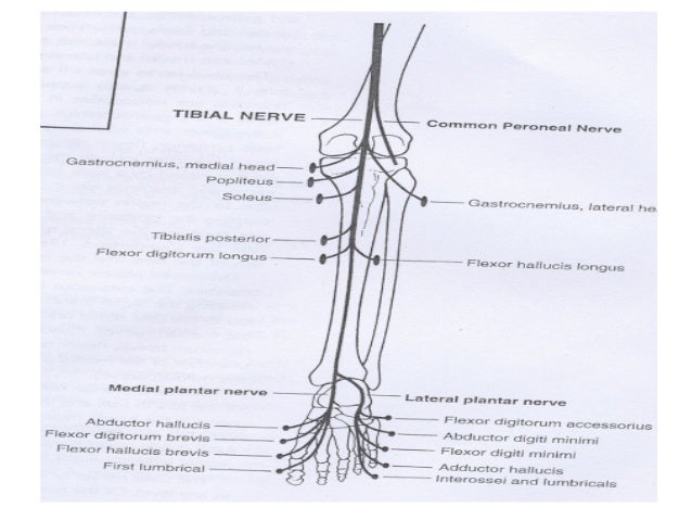 Anatomy Of Posterior Tibial Nerve By Im
 Leg Knee Anatomy
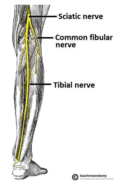 The Tibial Nerve Course Motor Sensory Teachmeanatomy
 Figure Posterior Tibial Artery Image Courtesy S Bhimji Md
Tibia Shinbone Shaft Fractures Orthoinfo Aaos
 Tibia Wikipedia
:max_bytes(150000):strip_icc()/posterior-tibial-tendonitis-2548561-5c77313dc9e77c000136a696.png) Posterior Tibial Tendonitis Signs And Treatment
Posterior Tibial Tendonitis Signs And Treatment

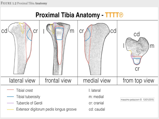 Tttt Tibial Tuberosity Transposition Tool And Technique
 Pediagenosis
 Figure 2 From Tibial Anatomy And Functional Axes Semantic
Tibia Shinbone Shaft Fractures Orthoinfo Aaos
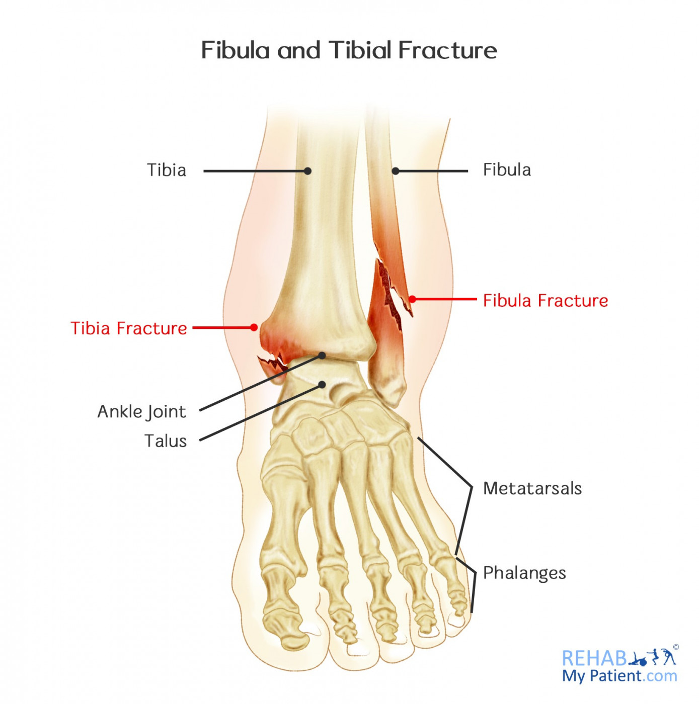 Fibula And Tibial Fracture Rehab My Patient
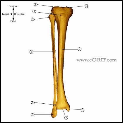 Tibial Shaft Anatomy Eorif
 Leg Knee Anatomy
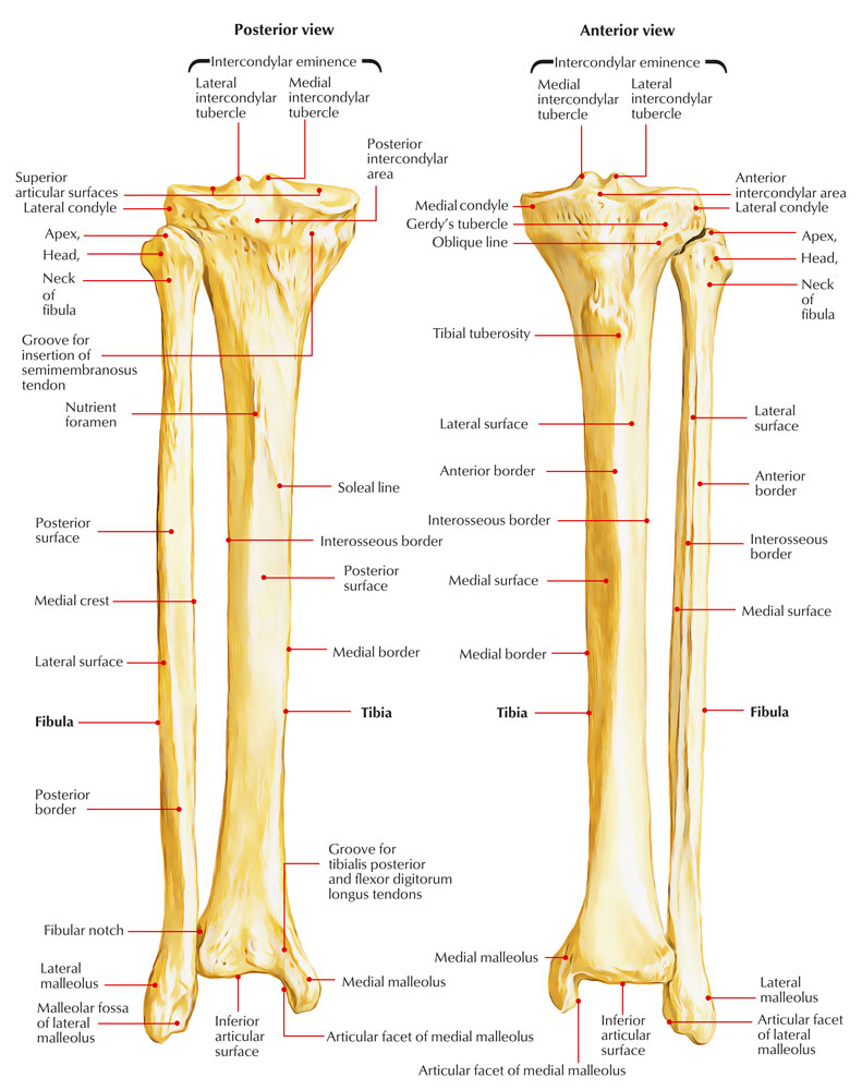
 Anatomy Of The Tibia Download Scientific Diagram
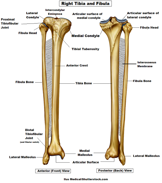 Tibia And Fibula Bone Anatomy

 Dorsalis Pedis Anterior Tibial Anatomy
 Understanding The Tibial Pedal Arterial Anatomy Practical
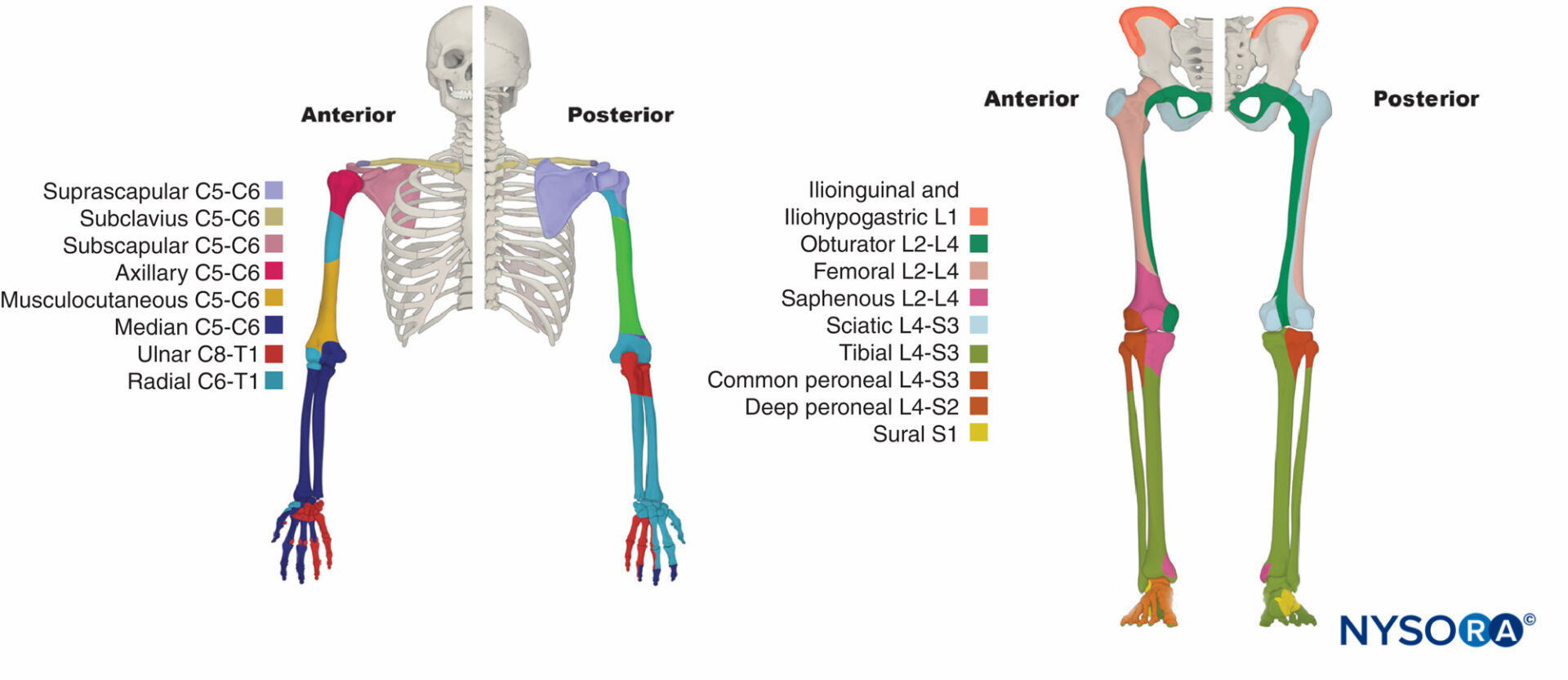
Functional Regional Anesthesia Anatomy Nysora
 Easy Notes On Tibia Shinbone Learn In Just 4 Minutes
 Shin Splints Medial Tibial Stress Syndrome Zion Physical
 Tibial Tubercle Fracture Pediatrics Orthobullets

 Chapter 36 Thigh The Big Picture Gross Anatomy
 Anterior Tibial Artery Wikipedia
 Tibial Plafond Fractures Trauma Orthobullets
 Right Posterior Tibial Vein The Anatomy Of The Veins Vis
 Posterior Tibial Vein Wikipedia


Posting Komentar
Posting Komentar