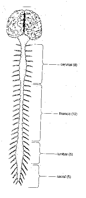
Rat brain pictures dorsal aspect of brain and rostral two ventral aspect of the brain and junction of segments of spinal cord. Paxinos george and charles watsonthe rat brain in stereotaxic coordinates.
 A Vascular Brain Anatomy Of The Rat Reproduced From 10
The rat brain is analyzed through stereotaxic localization of discrete brain areas and the subdivisions of many areas of rat brain are mapped using plates and diagrams.

Anatomy of rat brain. Thus after normalizing an individual image to the rat brain template the intracranial tissues could be conveniently extracted. A tool by matt gaidicamatt gaidica. Three principal strains are now commonly used for scientific study.
The is based on high resolution isotropic ex vivo t2 weighted mri and dti data acquired at the duke center for in vivo microscopy at resolutions of 39 μm and 78 μm respectively. The laboratory rat was developed from the norwegian rat rattus norvegicus by an american physiologist henry donaldson who started a breeding colony in 1906 at the wistar institute in philadelphia. The specimen is an 80 day old male sprague dawley rat.
The rat atlas is a three dimensional 3d computerized map of rat brain anatomy created with digital imaging techniques. Medulla with spinal cord. Access online via elsevier 2006.
Developed at the wistar institute. In addition an intracranial rat brain mask was formed from this canonical brain by assigning 1 for intracranial voxels and 0 for others as shown in figure 1b. The waxholm rat atlas is an open access volumetric atlas of the sprague dawley rat brain.
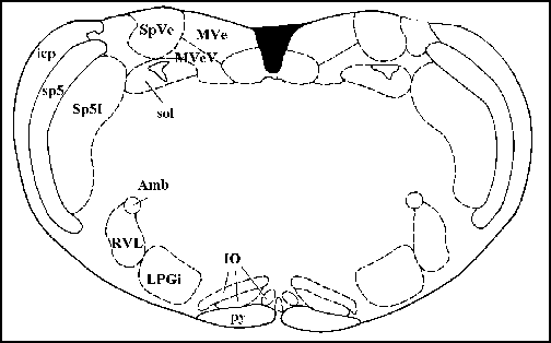
Photographs of sufficient magnification are included to permit investigators to judge for themselves the veracity of the atlas delineations. Electronic sharing and interactive use are benefits afforded by a digital format but the foremost advantage of this 3d map is its whole brain integrated representation of rat in situ neuroanatomy.
 Environmental Enrichment Wikipedia
 Enlarge
 Sucrose Intensity Coding And Decision Making In Rat
 Rat Brain Comparative Anatomy Model

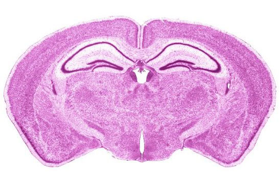
 A Color Atlas Of Sectional Anatomy Of The Rat Atlas
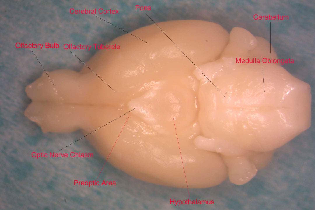
 Rat Brain Anatomy Slk Art
 Science Source Rat Brain Anatomy

 A Vascular Brain Anatomy Of The Rat Reproduced From 10

 Nervous System Sciencedirect
 Pdf Mitotic Activity In Adult Rat Brain Induced By
 Voltage Gated K Channel B Subunits Expression And
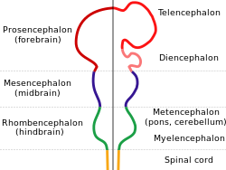
The Central Nervous System Biology 2e Openstax

 Figure 1 From Inhibitory Interneurons In The Piriform Cortex
 1 Anatomy Of The Hippocampal Formation A Schematic Rat
 Frontiers Three Dimensional Atlas System For Mouse And Rat
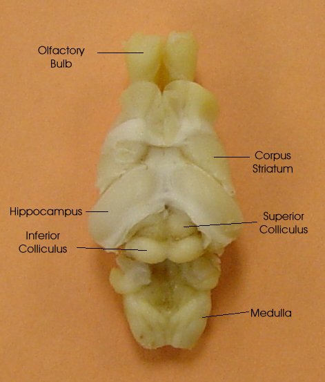
 Gross Neuro Anatomy


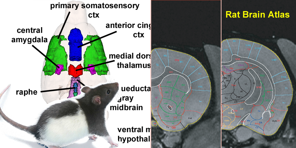
 Rat Brain Anatomy
 Brain Maps 4 0 Structure Of The Rat Brain An Open Access
 Brain Wikipedia
 Nervous System Sciencedirect


Posting Komentar
Posting Komentar