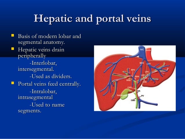
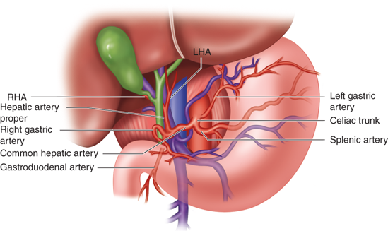
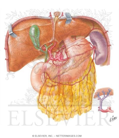
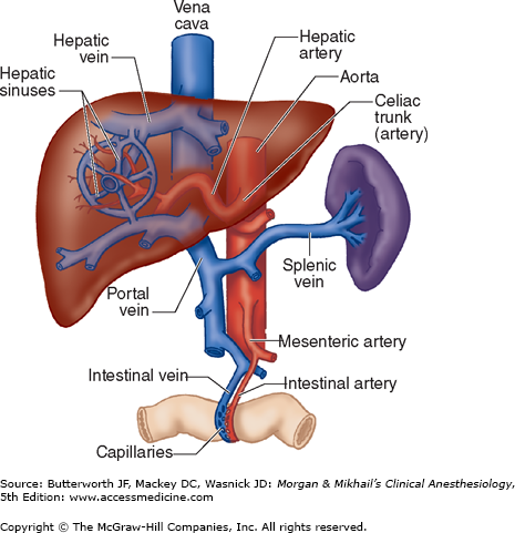
To prevent and decrease procedural complications it is important to identify variants of hepatic vascular anatomy before interventional radiologic procedures surgery and liver transplantation. The hepatic artery and portal vein furnish a double blood supply and venous drainage is handled by the hepatic veins.
 Liver Ultrasound
A vascular ultrasound of the liver is performed to help evaluate the liver and its network of blood vessels within the liver and entering and exiting the liver.
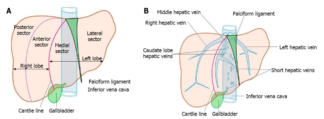
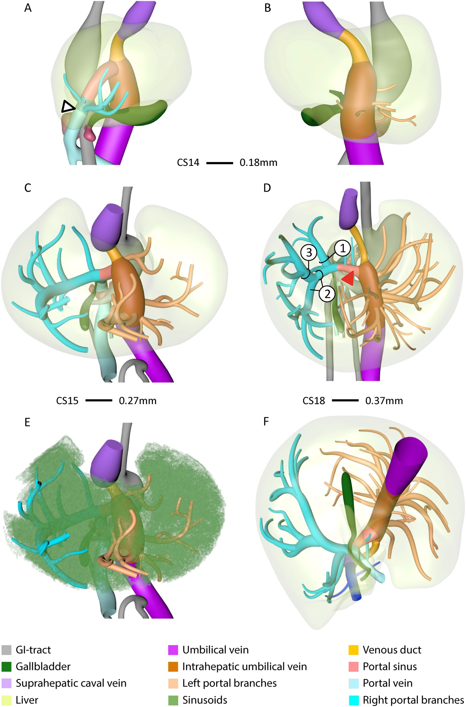
Liver vascular anatomy. At any point of time about 40 of blood volume in the liver is present in the large vessels and 60 of blood volume in the sinusoids. Knowledge of the vascular variants helps in selecting patients and in exploring alternative management options. It is pinkish brown in color with a soft consistency and is highly vascular and easily friable.
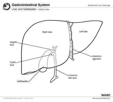
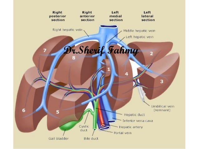
The liver has the general shape of a prism or wedge with its base to the right and its apex to the left see the image below. Confusion surrounds the nomenclature of liver anatomy. An accessory digestion gland the liver performs a wide range of functions.
The liver is a peritoneal organ positioned in the right upper quadrant of the abdomen. From the sinusoids blood flows into the central veins that drain via the hepatic veins hv into inferior vena cava ivc. The vascular anatomy of the liver is complex.
It is the largest visceral structure in the abdominal cavity and the largest gland in the human body. A refresher to prevent and decrease procedural complications it is important to identify variants of hepatic vascular anatomy before interventional. Request pdf liver vascular anatomy.
Including synthesis of bile glycogen storage and clotting factor production. Using vascular ultrasound can help physicians diagnose and review the outcome of treatments for various liver related problems and diseases.
 Liver Schwartz S Principles Of Surgery 10e
 Liver Anatomy Overview Gross Anatomy Microscopic Anatomy

 Linear Endoscopic Ultrasound Evaluation Of Hepatic Veins
 Arterial Variations And Collateral Supply Of Liver And
 Vascular Disorders Of The Liver And Splanchnic Circulation
Intrahepatic Vascular Anatomy In Rats And Mice Variations
 Chapter 32 Hepatic Physiology Anesthesia Morgan
 Full Text State Of The Art Cross Sectional Liver Imaging
 Frontiers Side Effects Of Yttrium 90 Radioembolization

 Human Liver Segments Role Of Cryptic Liver Lobes And
Vascular Ultrasound Of The Liver Cleveland Clinic
 Liver Irene S Film Critique
 Hepatic Vascular Anomalies Veterian Key
 The Liver Anatomy Of The Abdomen

 Gastrointestinal Radiology
 Startradiology
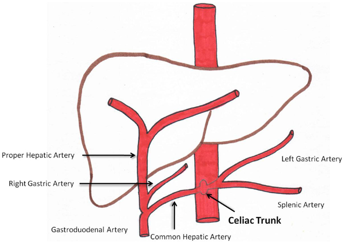
 Anatomy Of Liver Arteries For Interventional Radiology
![]() Hepatic Portal Vein Anatomy Function Clinical Points Kenhub
Hepatic Portal Vein Anatomy Function Clinical Points Kenhub
 Liver Wikipedia
 Anatomy Of Liver Arteries For Interventional Radiology
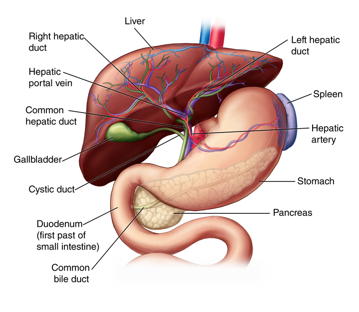
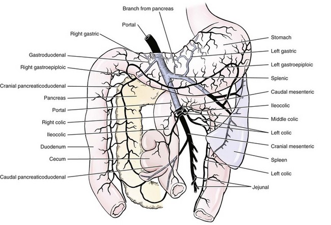
 The Pancreas Anatomy Duct System Vasculature


Posting Komentar
Posting Komentar