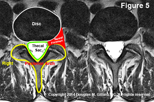
Mri of the lumbar spine a. The axial view also nicely visualizes the neural foramina posterior bony elements and paraspinal muscles.
 Thoracic Spine An Overview Sciencedirect Topics
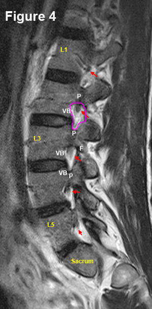
The lumbar spine consists of bones usually five vertebral bodies stacked on top of each other and separated by five discs.
Lumbar spine mri anatomy. Normal anatomy of the lumbar spine before deciphering the abnormalities listed in the lumbar mri report its important to understand the normal anatomy. On this view centered over the l4 l5 disk one can see well the cauda equina surrounded by csf note that csf is bright on t2 weighted images. To benefit all the functionalities of imaios we advise to keep the activation of all categories of cookies.
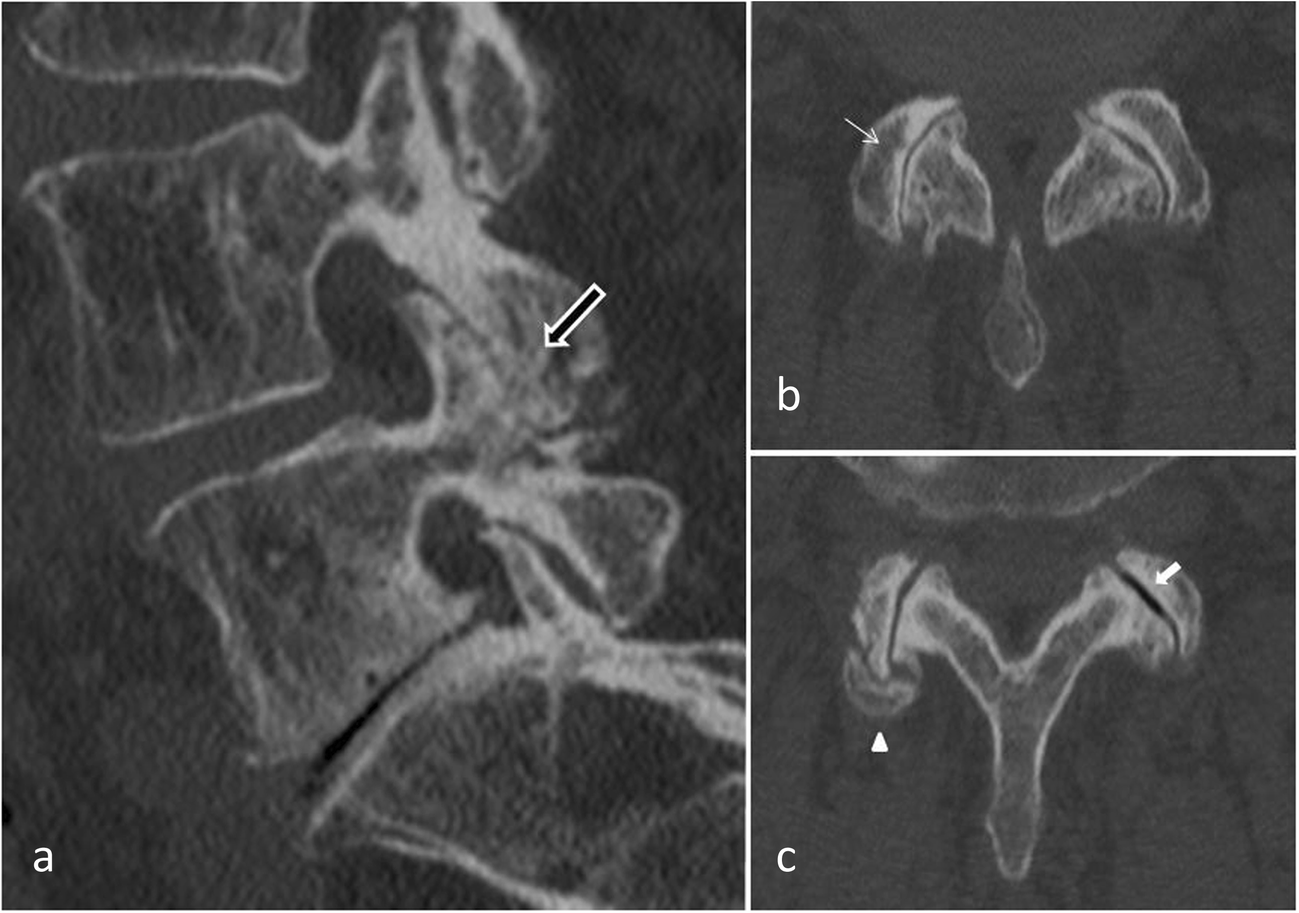
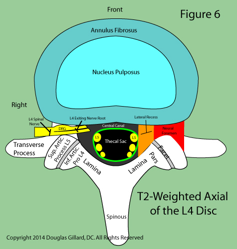
Lumbar spine anatomy on mri magnetic resonance imaging anatomy of the lumbar spine using cross sectional imaging mr t1 and t2 weighted. Axial mri of the lumbar spine t2 weighted image at the l4 level. Bony metastasis yellow arrow is seen involving the t12 vertebral body bpost contrast c sagittal t1wtd.
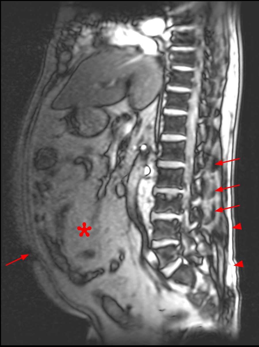
This mri lumbar spine cross sectional anatomy tool is absolutely free to use. This section of the website will explain large and minute details of lumbar spine sagittal cross sectional anatomy. General lumbar spine disc anatomy the lumbar spine low back is made up of five lumbar vertebrae backbones that are separated by five intervertebral discs discs the lowest of which sits on top of a large triangular shaped bone called the sacrum.
Sagittal coronal and transverse slices.
Mri Of The Lumbar Spine
 Lumbar Disc Herniation Spine Orthobullets
 Radiology Images
 The Radiology Assistant Spine Lumbar Disc Herniation
 Ultrasound Imaging Of The Lumbar Spine For Central Neuraxial
 Science Source Coronal Mri Of Lumbar Nerve Roots
 Lumbar Spine Anatomy On Mri Magnetic Resonance Imaging
 Mri Images Lumbar Spine T2 Fse Axial Mr Tip Com
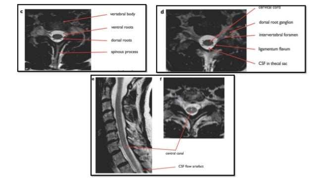
 Understanding Basic Mri Of The Spine
 Read Your Mri Basic Education From A World Renowned Spine
 Imaging The Cervical Thoracic And Lumbar Spine Radiology Key
 Mri Spine Anatomy
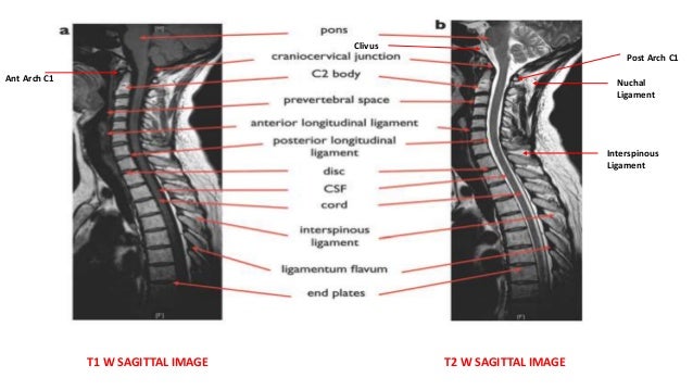
 What Is The Difference Between T1 And T2 Imaging In Mri
 Mri Spine Anatomy
 Sagittal T2 Weighted Image Of An Mri Of The Lumbar Spine
 Chirogeek Com
 Read Your Mri Basic Education From A World Renowned Spine
 Radiological Anatomy Of The Lumbar Spine X Ray Mri Ct Covered
 Pin By Nancy Kriskovich On Medical Lumbar Spinal Stenosis
 Mri Spine Anatomy
 Racgp Making Sense Of Mri Of The Lumbar Spine
 Spinal Sonography And Applications Of Ultrasound For Central

 Lumbar Spine Anatomy On Mri Magnetic Resonance Imaging

 Read Your Mri Basic Education From A World Renowned Spine
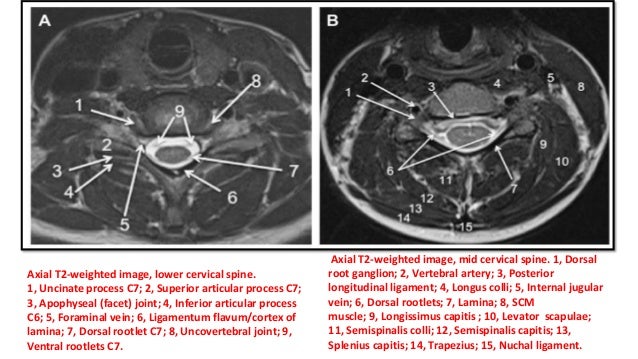
 Cervical Spine Mri
 Mri Spine Anatomy Free Mri Axial Cervical Spine Anatomy
Facet Joint Syndrome From Diagnosis To Interventional
 Cervical Spine Mri


Posting Komentar
Posting Komentar