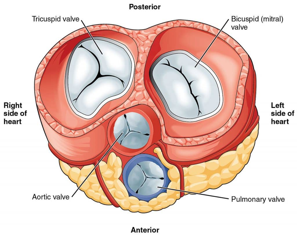
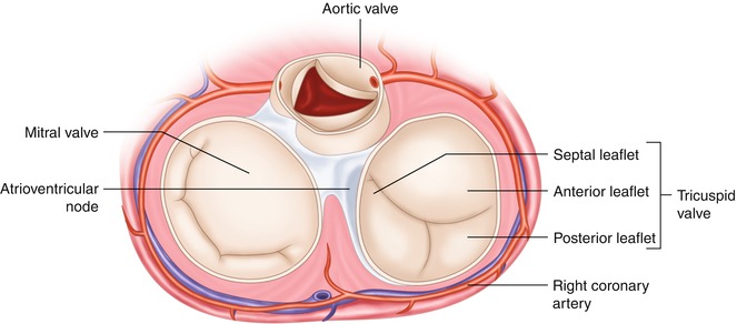
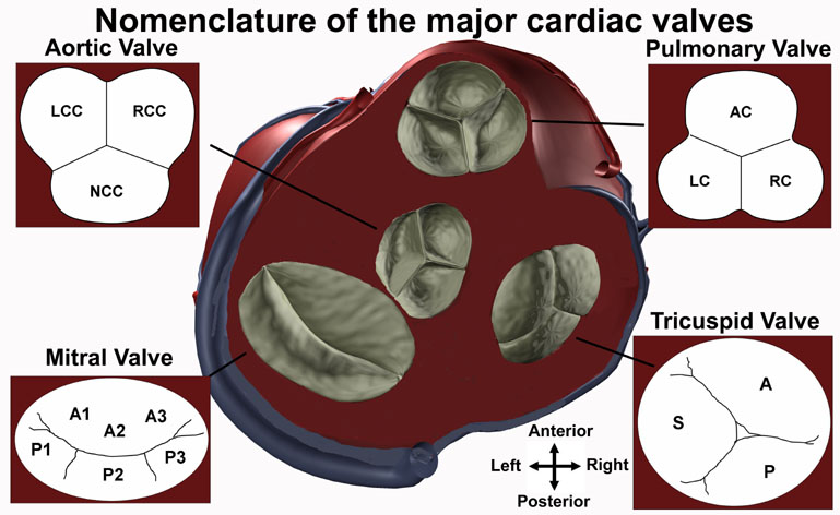
The tricuspid valve consists of three flaps or leaflets however cases are found when the tricuspid valve consists of only two or even four leaflets instead of the traditional three. The tricuspid valve lies within the right trigone.

The space in between the septal insertion of the tricuspid valve and the septal insertion of the anterior leaflet of mitral valve belongs to the membranous septum that separates the left ventricle from the right atrium.
Tricuspid valve anatomy. An appreciation of the complex and variable anatomy of the tricuspid valve is essential to unraveling the pathophysiology of tricuspid regurgitation. It is the atrioventricular valve that allows blood to flow from the right atrium to the right ventricle. Then blood exits the heart via the pulmonary artery.
Anatomy of the tricuspid valve. The tricuspid valve complex consists of the annulus leaflets right ventricle papillary muscles and chordae tendinae. Right atrioventricular valve tricuspid valve these are large veins that transport deoxygenated blood from the body back to the heart.
A greater appreciation of normal and abnormal anatomy is important as new methods of treating the tricuspid regurgitation are developed. It opens during diastole and closes during systole. The tricuspid valve tv is a complex structure.
B the relevant anatomy shown from the front view. The tricuspid valve lies between the right atrium and the right ventricle and is placed in a more apical position than the mitral valve. The tricuspid valve has an area of 4 to 6 cm square and is located between the right atrium and the right ventricle of the heart.
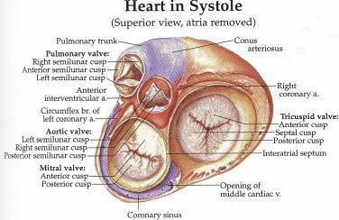
The valve has anterior posterior and septal leaflets cusps the bases of which attached around the av orifice to the fibrous ring fibrous skeleton of the heart. Blood collects within the right atrium and it must flow through the tricuspid valve in order to enter the right ventricle. The tricuspid valve functions as a one way valve that closes during ventricular systole to prevent regurgitation of blood from the right ventricle back into the right atrium.
A the anatomy of the tricuspid valve and adjacent structures from a surgical view. Anatomy of the tricuspid valve. The tricuspid valve anatomy shows greater variability than the anatomy of the mitral valve.
In normal heart the tricuspid valve is located slightly closer to the apex than the mitral valve. The red dotted lines show the direction of dilation of various structures in the setting of secondary tricuspid regurgitation. Unlike the aortic and mitral valve it is not possible to visualize all tv leaflets simultaneously in one cross sectional view by standard two dimensional echocardiography 2de either transthoracic or transesophageal due to the position of tv in the far field.
It opens during ventricular diastole allowing blood to flow from the right atrium into the right ventricle.
State Of The Art Review Of Echocardiographic Imaging In The
 Cardiac Interventions Today Transcatheter Tricuspid Valve
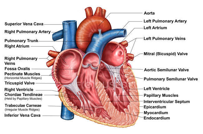
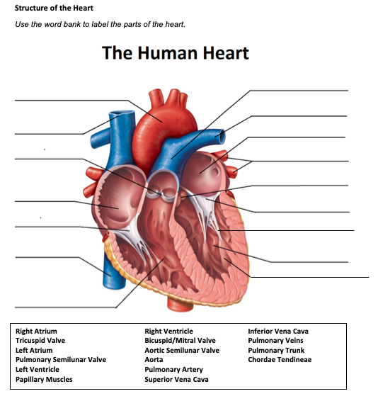 Solved Structure Of The Heart Use The Word Bank To Label
 Auscultating Heart Soundsin The Following Procedure Yo
 Anatomy Of The Tricuspid Valve
 Heart Valve Anatomy Tricuspid Valve Png Clipart Anatomy
 Heart Valves Function And Anatomy How The Heart Valves Work
 Heart Valves And Fibrous Skeleton Mitral Valve Tricuspid
 Anatomy Of Heart Interior With Labels Cross Section Blood
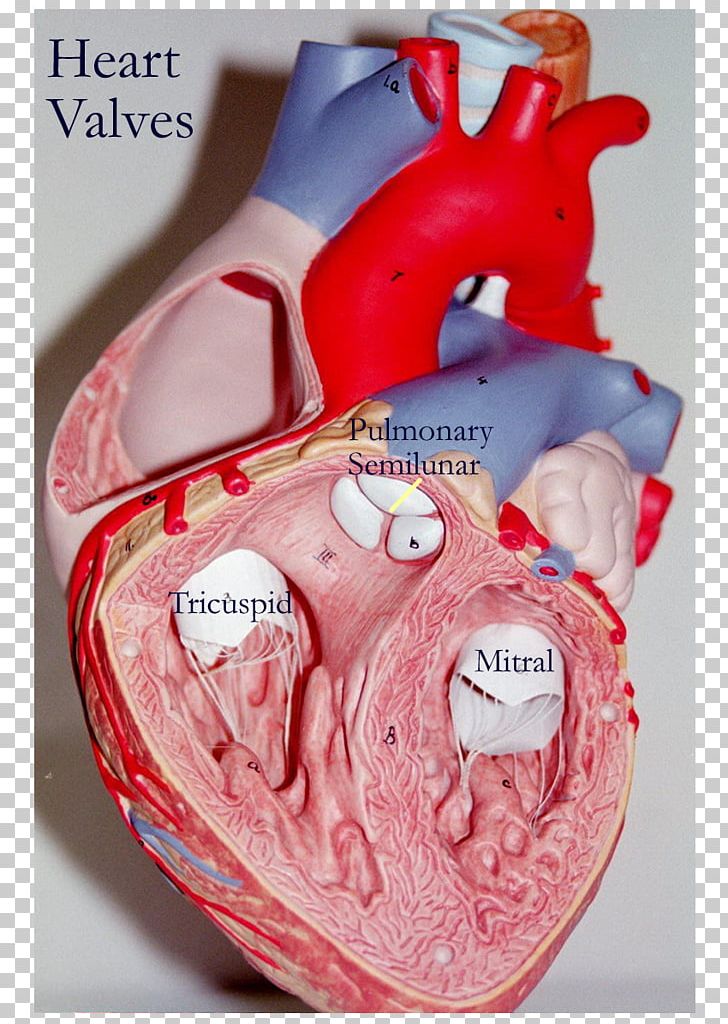
 Heart Model With Valves
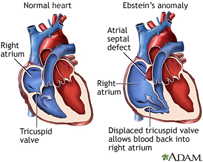
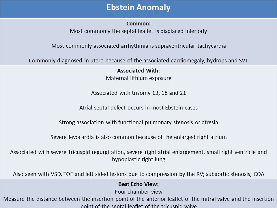
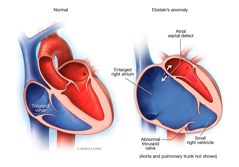 Cone Reconstruction For Ebstein Anomaly Mayo Clinic
 The Heart Valves Tricuspid Aortic Mitral Pulmonary
 Anatomy Of Tricuspid Valve
 Pin By Melissa Blaker On Cardiac Surgery Tricuspid Valve
 Anatomy And Physiology Of The Tricuspid Valve Sciencedirect
 Tricuspid Valve Anatomy Overview Gross Anatomy
 Isolated Tricuspid Regurgitation Outcomes And Therapeutic
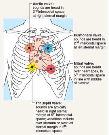
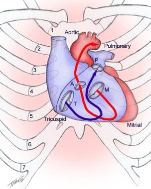
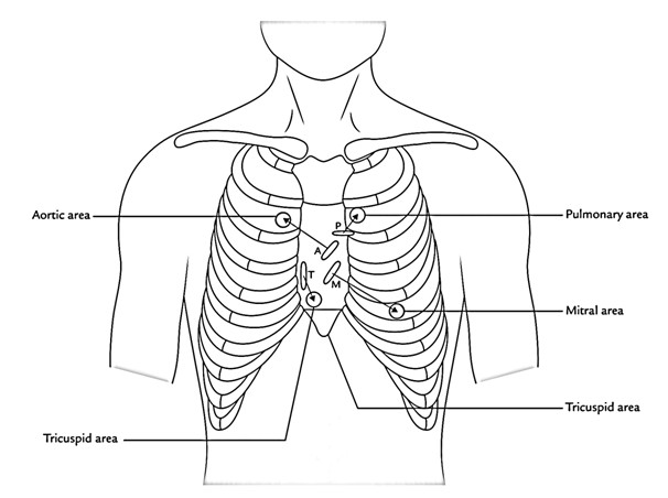 Surface Markings Of The Cardiac Valves And Auscultatory
 Cardiac Interventions Today Echocardiography For Tricuspid
 Anatomy Of The Tricuspid Valve And Pathophysiology Of
 Anatomy Of The Normal Tricuspid Valve Showing Orientation Of
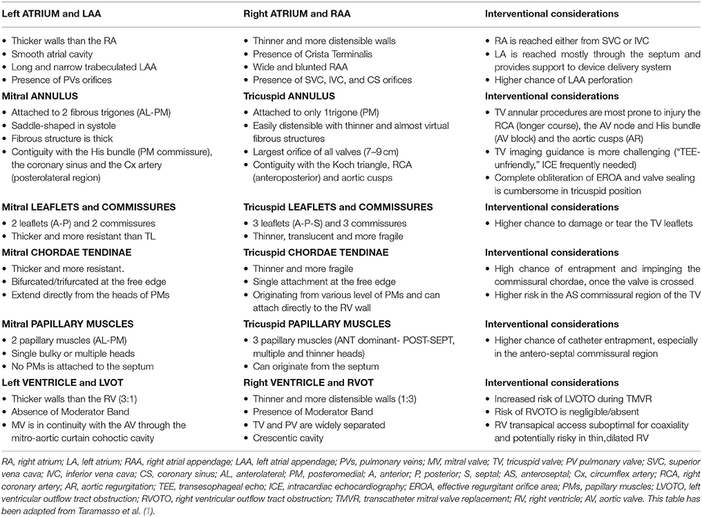
Frontiers Comparative Anatomy Of Mitral And Tricuspid
 Diagnosis And Treatment Of Tricuspid Valve Disease Current
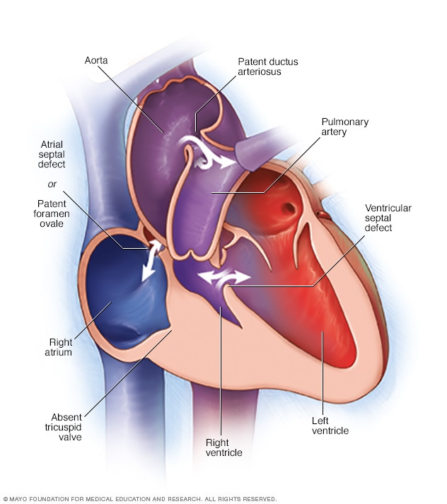 Tricuspid Atresia Symptoms And Causes Mayo Clinic
 Ultrasound Registry Review Valvular Abnormalities


Posting Komentar
Posting Komentar