Basal forebrain on ct and mr images the basal forebrain is a rather featureless region on the ventral surface of the brain. 6 frontal bone 27 occipital bone 32 optic nerve 37 basilar artery 40 hemisphere of cerebellum 43 frontal sinus 45 sigmoid sinus 46 internal carotid artery 47 sphenoid bone 49 medulla oblongata 50 external auditory meatus 51 spinal central canal.
 Ct Neck Brain Angiography
Anatomy of the head on a cranial ct scan.
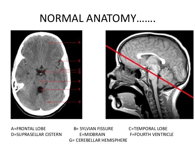
Ct brain anatomy. Brain bones of skull paranasal sinuses. Ct images of the brain are conventionally viewed from below as if looking up into the top of the head. The anterior part of the head is at the top of the image.
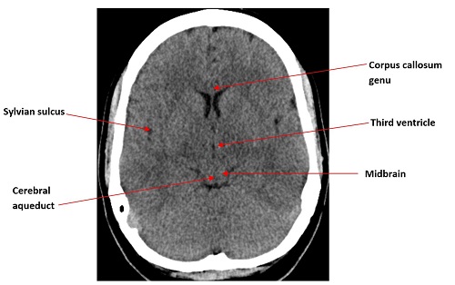
Non contrast axial ct head. This article lists a series of labeled imaging anatomy cases by system and modality. Angiogram axial ct head.
Ct brain image orientation. Coronal brain ct. Angiogram coronal ct head.
This lecture is a part of basic radiologic anatomy series. This means that the right side of the brain is on the left side of the viewer. Interactive anatomy atlas.
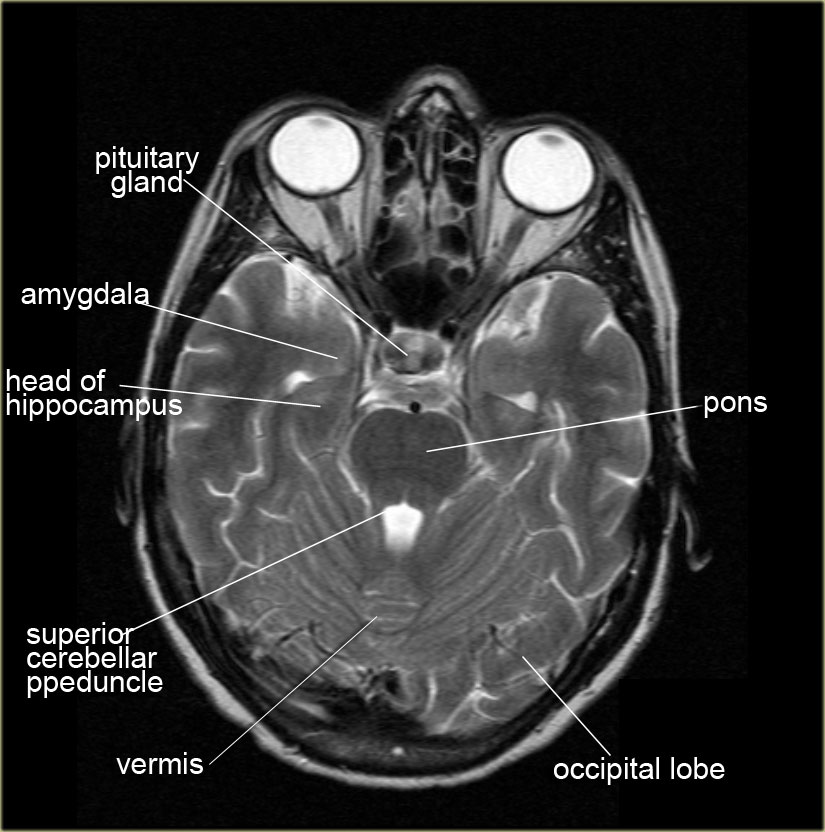
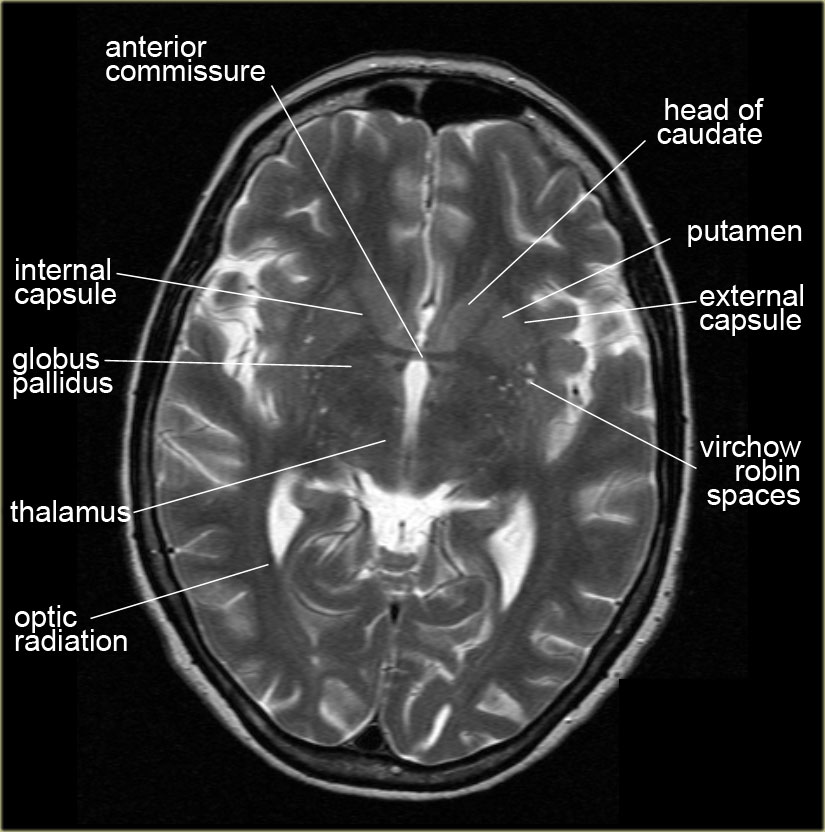
It contains both regions of gray and white matter in a heterogeneous fashion that are best appreciated on t1 and t2 weighted mr coronal images. Brain and face ct. Brain bones of cranium sinuses of the face.
The lecture discussing the basic ct anatomy of the brain. Non contrast sagittal ct head. Non contrast coronal ct head.
Cross sectionnal anatomy of the head on a cranial ct scan. Anatomy ct axial brain form no 18. Head ct anatomy normal anatomy 1.
 The Radiology Assistant Brain Anatomy
![]() Medical Imaging And Radiological Anatomy X Ray Ct Mri
Medical Imaging And Radiological Anatomy X Ray Ct Mri
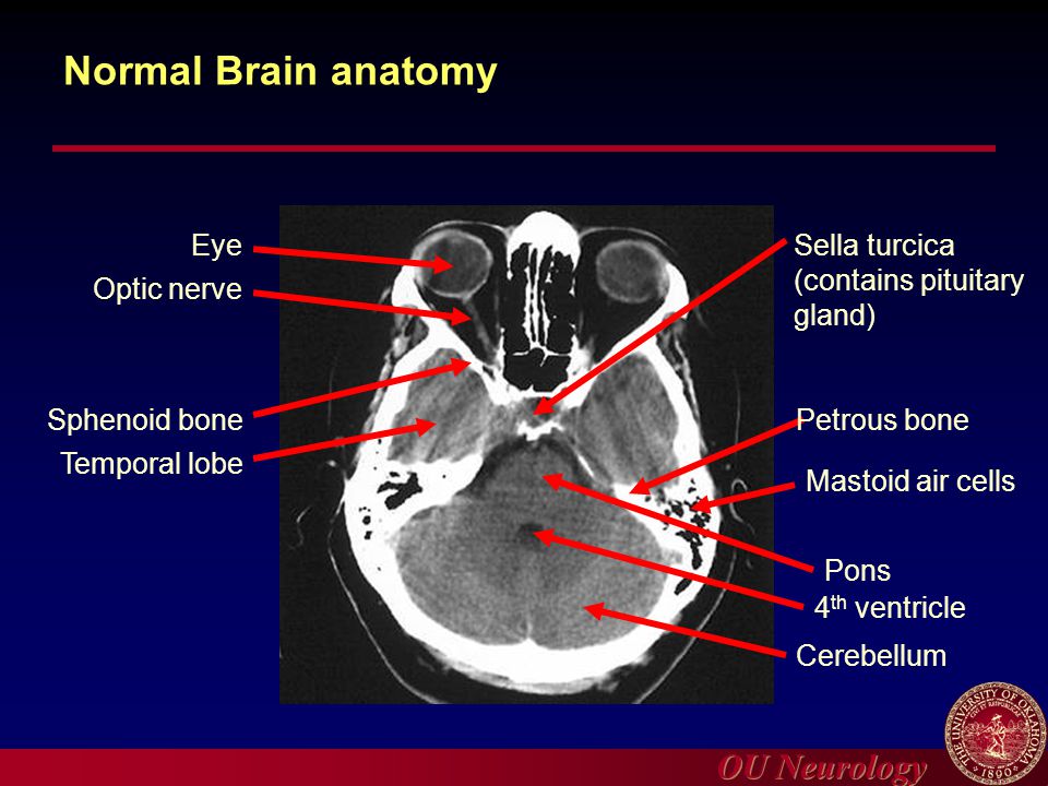
 Brain Ct Neurologyneeds Com
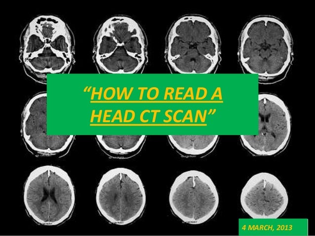
 Introduction To Head Ct Imaging Ppt Video Online Download
 Brain And Face Ct Interactive Anatomy Atlas
 Basics Of Ct Head
 Brain And Face Ct Interactive Anatomy Atlas
 Axial View Of A Head Computed Tomography Ct Scan Of Pineal
 The Radiology Assistant Brain Anatomy
 Can T Miss Findings On Noncontrast Head Ct Slideshow
 How To Easily Tell The Difference Between Mri And Ct Scan
 Radiology Basics Head Anatomy
 Ct Brain For Android Apk Download
 Brain Imaging
 Brain Lobes Annotated Mri Radiology Case Radiopaedia Org
 Mri Anatomy Free Mri Axial Brain Anatomy
Basics Of Ct Head

![]() Axial Transverse View Computed Tomography Ct Stock Photo
Axial Transverse View Computed Tomography Ct Stock Photo
 Cross Sectional Imaging Anatomy Thorax Youtube This Video
 Ct Brain Anatomy Basal Ganglia Google Search Mri Brain
 Confluence Mobil Tum Wiki
 Head Ct Scan Procedure Radtechonduty


Posting Komentar
Posting Komentar