It also includes the joints of the hip stifle hock fetlock pastern and coffin. Equine anatomy refers to the gross and microscopic anatomy of horses and other equids including donkeys and zebras.
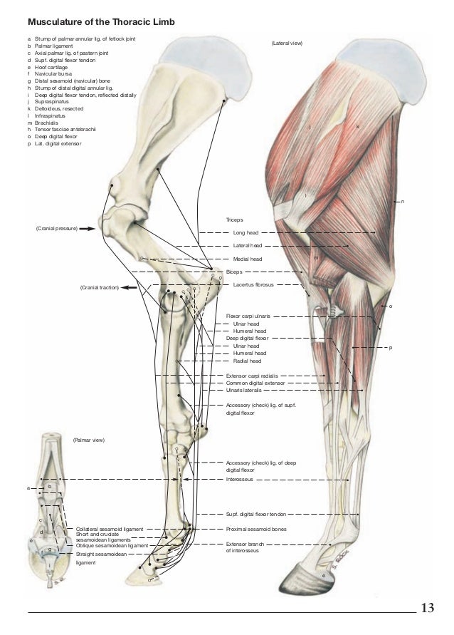
 The Topographical Anatomy Of The Limbs Of The Horse Page 43
While all anatomical features of equids are described in the same terms as for other animals by the international committee on veterinary gross anatomical nomenclature in the book nomina anatomica veterinaria there are many horse specific colloquial terms used by equestrians.
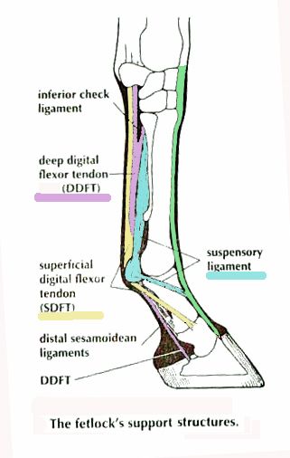
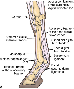
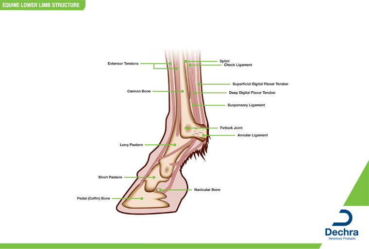
Horse limb anatomy. The suspensory apparatus which carries much of the weight prevents overextension of the joint and absorbs shock and the stay apparatus which locks major joints in the limbs allowing horses to remain standing while relaxed or asleep. Limbs of the horse the limbs of the horse are structures made of many bones joints muscles tendons and ligaments that support the weight of the horses body. They include two apparatuses.
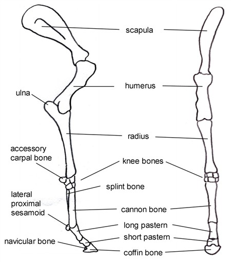
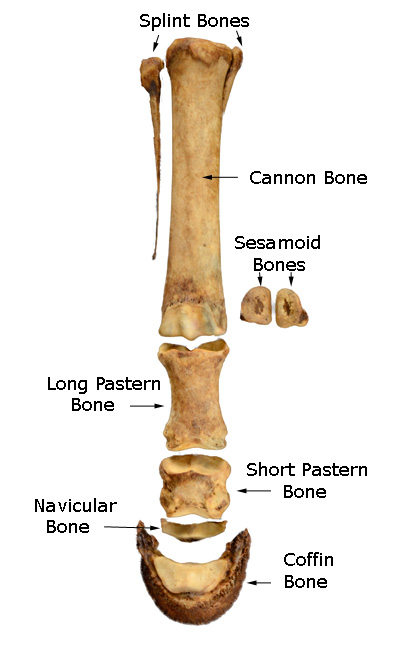
Select a body part and angle on the left then select the type of image from the top menu. In the young horse they are separate but often the splint bones fuse to the cannon bone. Offering anatomy charts and freeze dried and skeletal models of horse limbs.
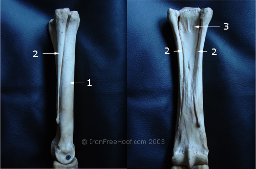
There are three bones between the knee and fetlock joints there are only three bones in this region. This is the same cannon bone in each photo. The limbs of the horse are structures made of dozens of bones joints muscles tendons and ligaments that support the weight of the equine body.
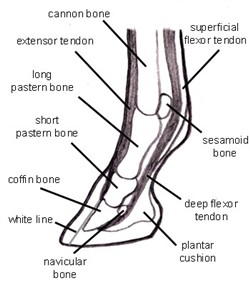
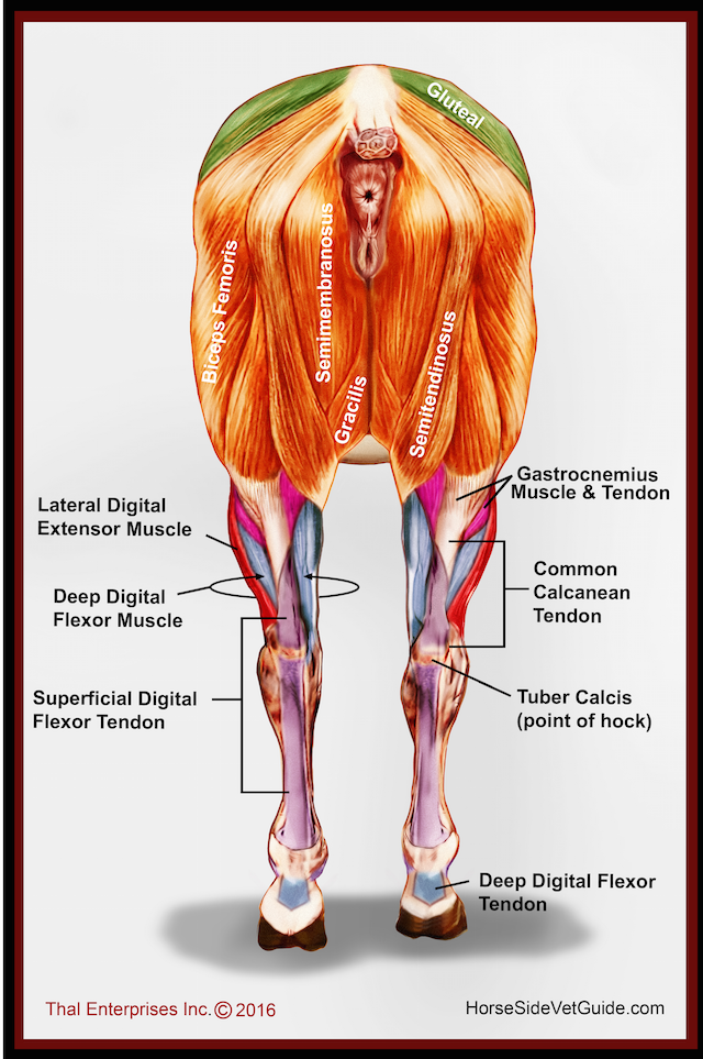
Equine rear leg bones and function the horse leg anatomy in the rear includes the bones of the pelvis the ilium ischium and pubic bones femur tibia fibula metatarsus and the phalanxes. It is very common for the splint bones to fuse with the cannon. These diagrams should explain and show you some of the basics.
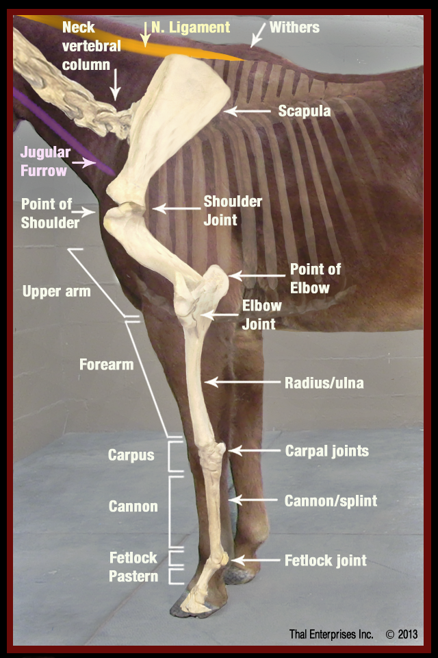
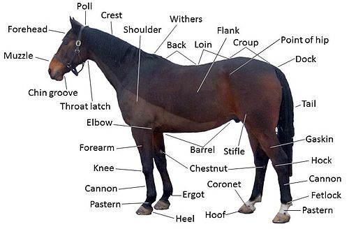
For something more basic or to use as a reference point check out this page for a downloadable labeled basic body parts diagram. That way if you need to talk to a vet or do a correct drawing youll have a solid foundation. It also includes the joints of the hip stifle hock fetlock pastern and coffin.
In the young horse there are three separate bones in the cannon region of the leg. Horse anatomy diagrams with printable pdfs directional terms skeletal and muscle introduction. In this example one splint bone is fused and the other is not.
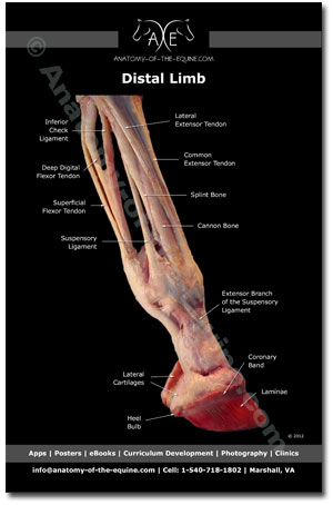
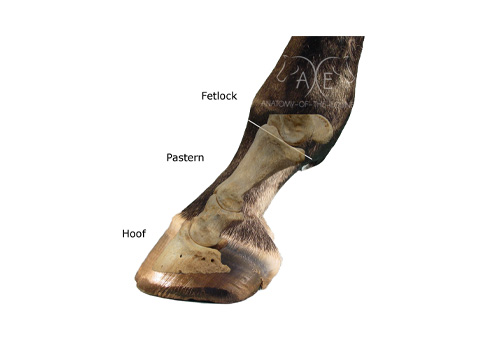
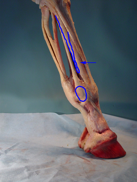
The horse leg anatomy in the rear includes the bones of the pelvis the ilium ischium and pubic bones femur tibia fibula metatarsus and the phalanxes. The equine distal limb visit my other website anatomy of the equine to see more anatomy of the lower leg. The limbs play a major role in the movement of the horse with the legs performing the functions of absorbing impact bearing weight and providing thrust.
 Anatomy Of The Horse
 Horse Limb Anatomy Diagram Quizlet
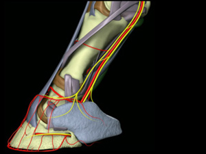
 Regional Anesthesia In Equine Lameness Musculoskeletal
 Horse Leg Vs Human Hand Horse Anatomy Horse Human
 Vitals Anatomy Horse Side Vet Guide
 The Topographical Anatomy Of The Limbs Of The Horse Horses
 Horse Hoof Anatomy Picture Click Quiz By Missdianap
 Horse Bones Everything You Need To Know To Get Started
 Limbs Of The Horse Wikipedia
 The Biomechanics Of The Equine Limb And Its Effect On
 Limbs Of The Horse Wikipedia
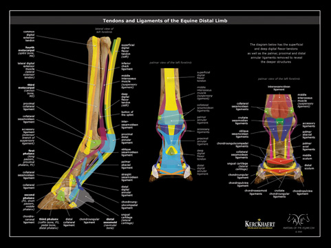
 The Glass Horse Elements Of The Equine Distal Limb
 Tendon Ligament Bone And Cartilage Anatomy Physiology
 Equine Anatomy Wikipedia
 Disorders Of The Shoulder And Elbow In Horses Horse Owners
 Ivala Learn 3d Veterinary Anatomy Clinical Learning Content
 Equine Forelimb Muscles Scan 3d Veterinary Anatomy Learning Ivala
 Vitals Anatomy Horse Side Vet Guide
 Ivala Learn 3d Veterinary Anatomy Clinical Learning Content
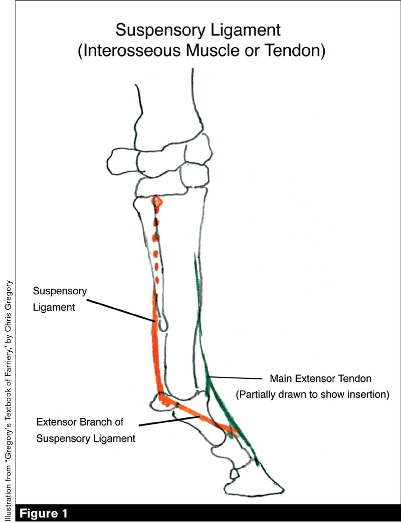 Increased Knowledge Of The Equine Anatomy Can Help Farriers
The Amazing Horse Hoof Think Like A Horse Rick Gore


Posting Komentar
Posting Komentar