Each is a glossy white complex tissue comprised of cells specialized extracellular matrix ecm molecules and region specific innervation and vascularization. The medial meniscus is often injured when the knee is twisted or sprained with sudden force.
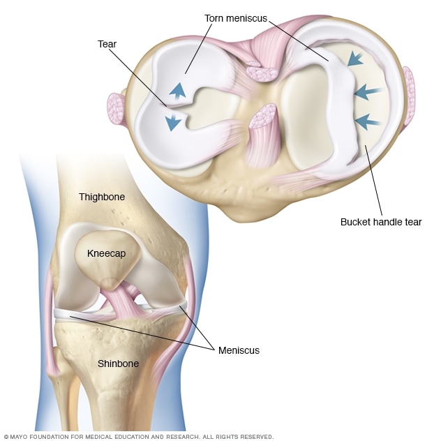
The meniscus is a c shaped piece of tough rubbery cartilage that acts as a shock absorber between your shinbone and thighbone.
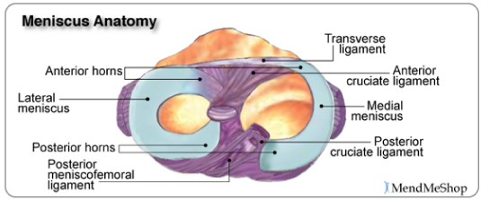
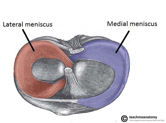
Knee meniscus anatomy. A basic meniscus definition is a crescent shaped fibrous cartilage between the bones at certain joints especially in the knees the knee is made up of the femur the tibia and the patella bones. There are two knee menisci in each joint. Anatomy and function of the menisci.
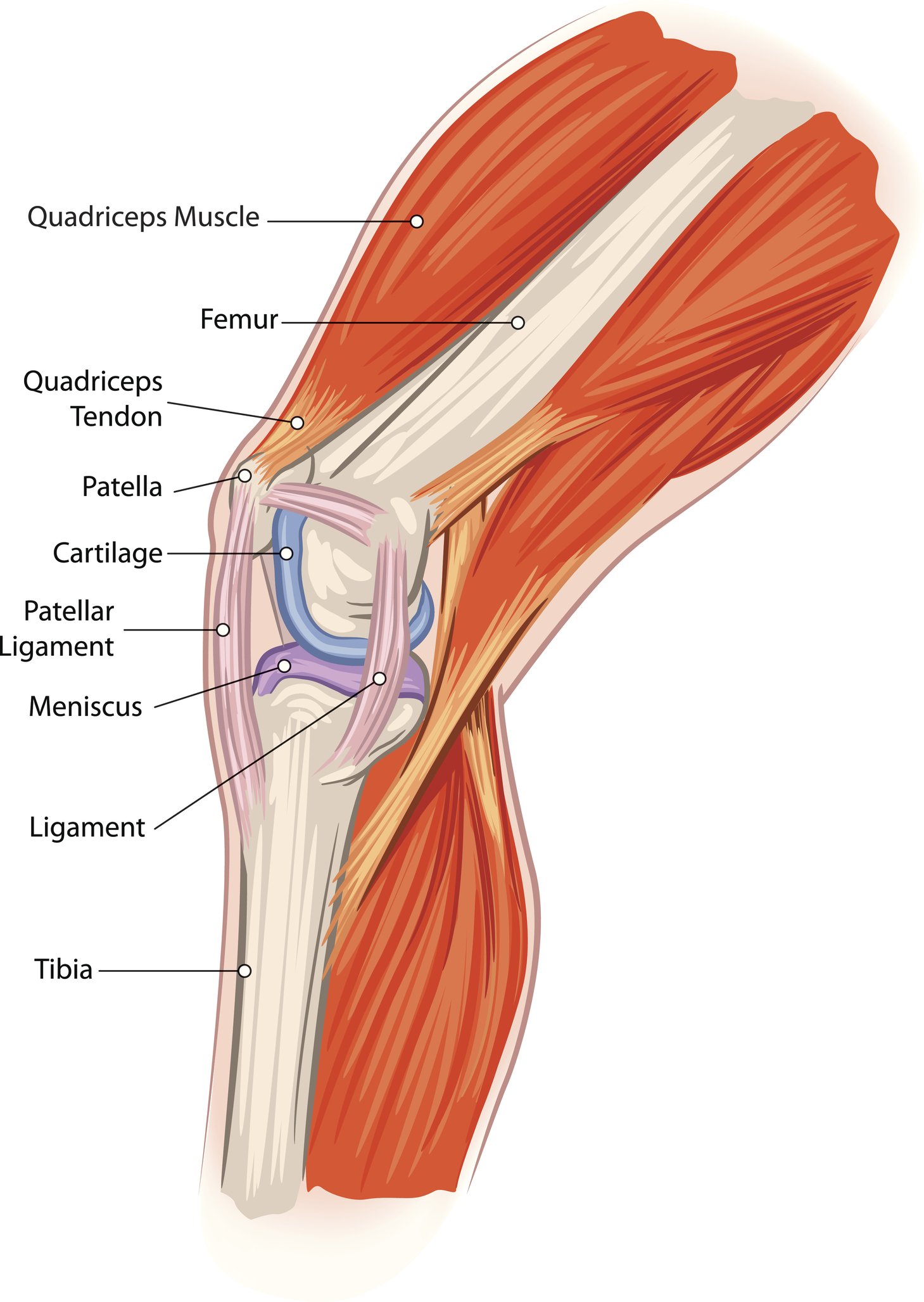
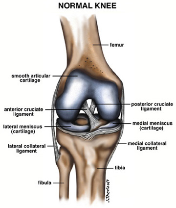
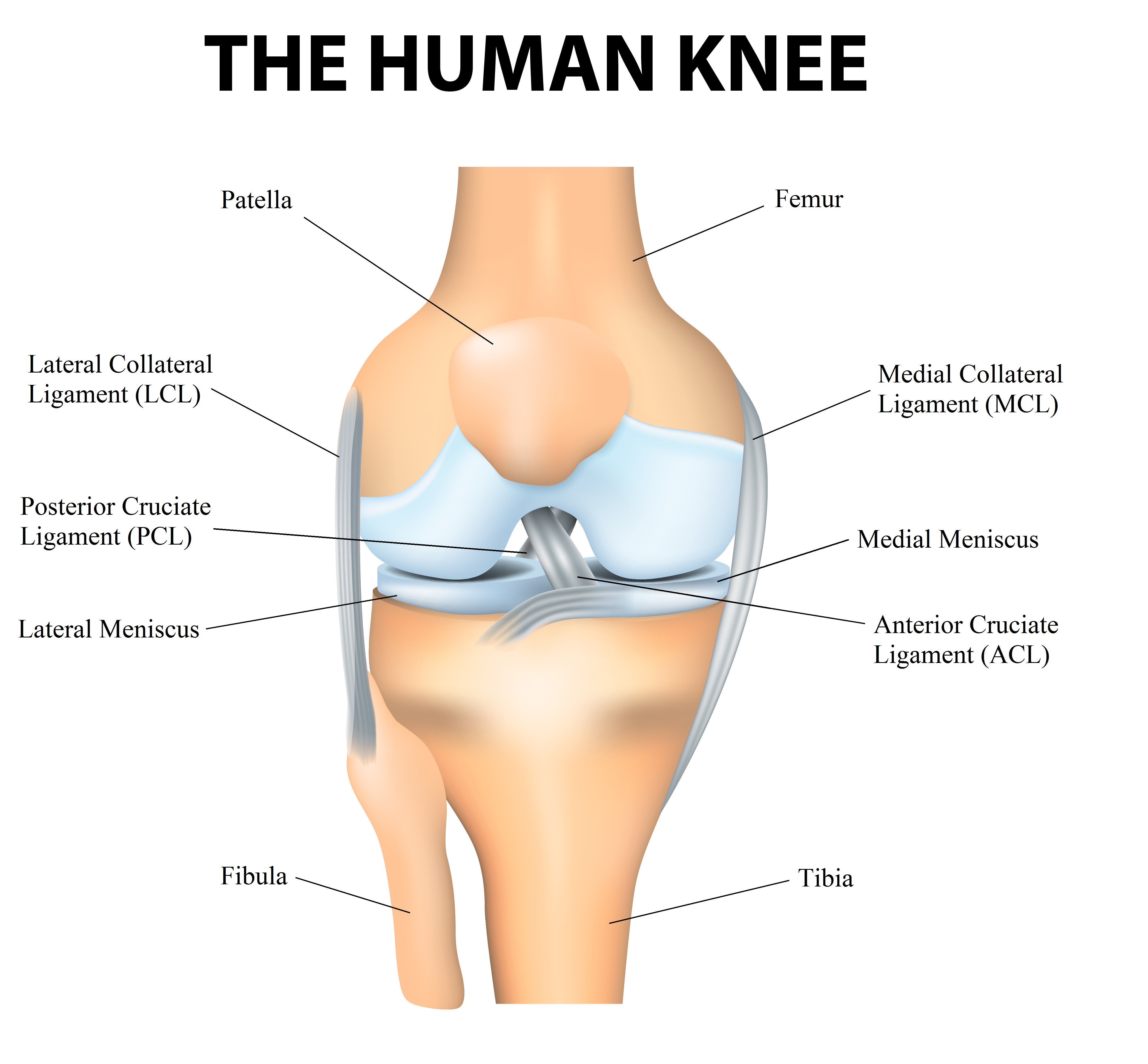
It is not the bones inside the knee that provides stability instead it is the soft tissue tendons ligaments muscles menisci that hold the femur thigh bone the tibia shinbone the fibula the slender bone in the lower leg and the patella kneecap together at the joint. In most of our joints including the knee there is a layer of articular cartilage which is made of collagen and chondroitin. The knee meniscus is a special layer of extra cartilage that lines the knee joint.
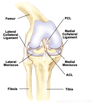
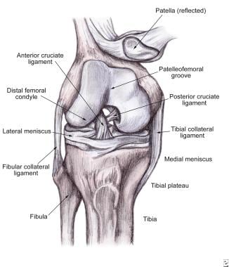
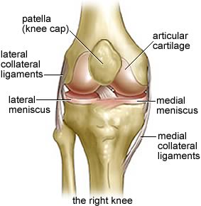
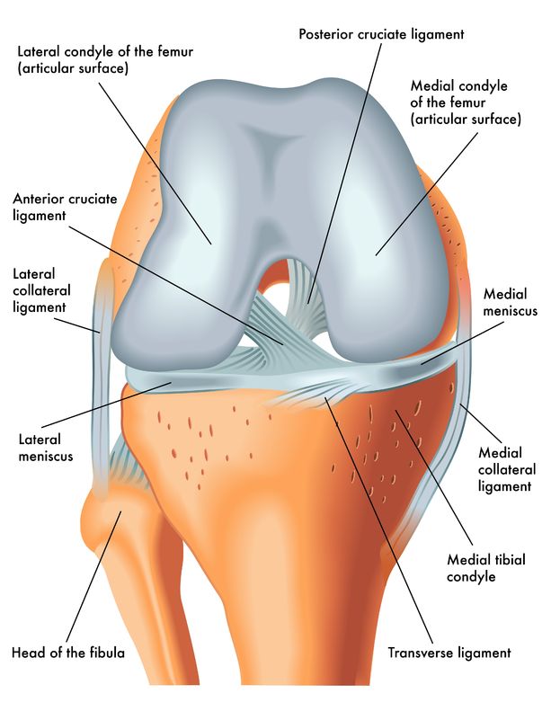
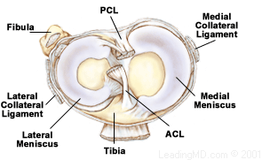
Pain swelling and warmth in any of the bursae of the knee. The knee joint contains the meniscus structure comprised of both a medial and a lateral component situated between the corresponding femoral condyle and tibial plateau figure 1. The articular capsule at the knee joint is thin and in some areas is incomplete but is strengthened by various ligaments and tendons of associated muscles.
The menisci are described as having a central body with anterior and posterior horns. Collection of fluid in the back of the knee. Ligaments are tough fibrous connective tissues which link bone to bone made of collagen.
In knee joint anatomy knee ligaments are the main stabilising structures of the knee preventing excessive movements and instability. Each meniscus has a differing shape size and attachments. Anatomy of the meniscus and knee.
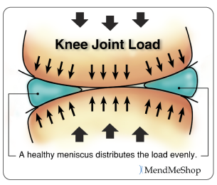
They are concave on the top and flat on the bottom articulating with the tibia. A torn meniscus is one of the most common knee injuries. It provides a smooth surface over the bones.
Bursitis often occurs from overuse or injury. External rotation rotating the knee outward puts the most strain on the meniscus while inward internal rotation is the least strenuous. It can be torn if you suddenly twist your knee while bearing weight on it.
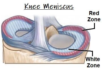
In cross section they have a triangular bow tie shape being thicker peripherally and thinning to a free edge centrally. What is a meniscus. Its job is to cushion the joint and transfer forces between the tibia and femur bones.
Meniscus anatomy the menisci of the knee are two pads of fibrocartilaginous tissue which serve to disperse friction in the knee joint between the lower leg tibia and the thigh femur. Ligaments hold the bones of the knees together. It is less mobile than the lateral meniscus because it is firmly attached to the tibial collateral ligament.
They are attached to the small depressions fossae.

 Soft Tissue Knee Injury Practice Essentials Background
 What Is A Meniscus
 The Knee Meniscal Injuries
 Pin On Yoga
 Knee Pain Meniscus Tears Colorado Springs Sports Doc
 The Knee Anatomy Injuries Treatment And Rehabilitation
 Meniscal Repair Physiopedia
 Arthroscopy Pinnacle Orthopaedics
 Meniscal Tears Classification Surgical Repair

 Torn Meniscus Anatomy And Causes Video Jeffrey H Berg
 Knee Meniscus Cartilage Knee Pain Explained
 Menicus Injuries United States The Orthopedic Center
 Torn Meniscus Symptoms And Causes Mayo Clinic
 Atro Medical Meniscus Vervanging Replacement Atro Medical
 4 Common Causes Of Knee Pain
 Physical Therapy To Treat Torn Meniscus Comparable To
 Regeneration Of Knee Joint Menisci Methods Review Bio

 Lateral Meniscus Physiopedia

Meniscus Injuries


Posting Komentar
Posting Komentar