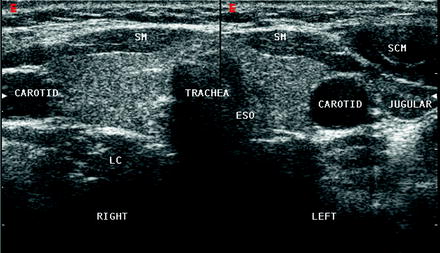
The visceral space contains the thyroid parathyroid glands larynx hypopharynx the cervical trachea and esophagus the recurrent laryngeal nerve. Optimal positioning and exposure of the neck for ultrasound of the thyroid and parathyroid glands a b and lateral neck for lymph node examination and mapping c.
 Startradiology
Ultrasonographic examination of head and neck pathology is cost efficient non irradiating and permits fast follow up with serial examination of the lesions.
Neck ultrasound anatomy. While the vast majority of patients are supine on the exam table with a pillow supporting the shoulders to allow gentle neck extension keep in mind that some patients have beautiful anatomy d that allows ultrasound exam even in a sitting position. Specifically it is an effective clinical tool to evaluate head and neck anatomy and pathology. The infrahyoid region of the neck includes the visceral anterior cervical posterior cervical carotid retropharyngeal and perivertebral spaces.
Find out more from alaska family sonograms. Ultrasonography is highly useful for many disease processes and many organ systems. Anterior neck anatomy false vocal cords true vocal cords paraglottic fat.
The infrahyoid region of the neck includes the visceral anterior cervical posterior cervical carotid retropharyngeal and perivertebral spaces. Furthermore one can perform an initial tnm staging of the case prior to other expensive imaging studies such as ct and mri. The visceral space contains the thyroid parathyroid glands larynx hypopharynx the cervical trachea and esophagus the recurrent laryngeal nerve.
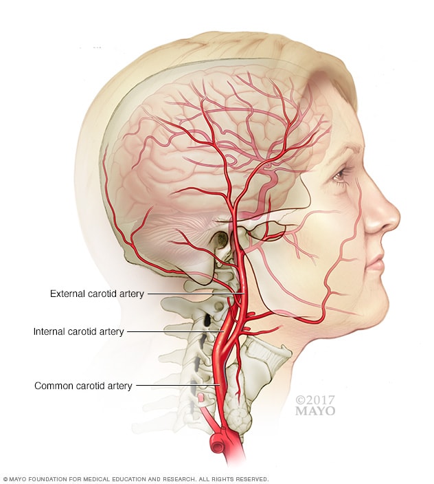
It does not use ionizing radiation. A common neck ultrasound is ultrasound of the thyroid which uses sound waves to produce pictures of the thyroid gland within the neck. A neck ultrasound is performed to diagnose potential problems of the thyroid lymph nodes and carotid arteries.
It can play an.
 Topographical Features Of The Vagal Nerve At The Cervical
 What Is An Anatomy Ultrasound During Pregnancy Babymed Com
Normal Neck Anatomy And Method Of Performing Ultrasound
Surveillance Neck Sonography After Thyroidectomy For
 Normal Neck Ultrasound Radiology Case Radiopaedia Org
 The Radiology Assistant Infrahyoid Neck
 Neck Ultrasound


 Carotid Ultrasound Mayo Clinic
 Computed Tomography Of The Thyroid Wikipedia
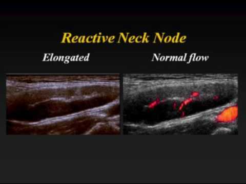 Ultrasound Of Cervical Lymph Nodes
 Chapter 4 Ultrasound Of The Neck Thyroid And Parathyroid
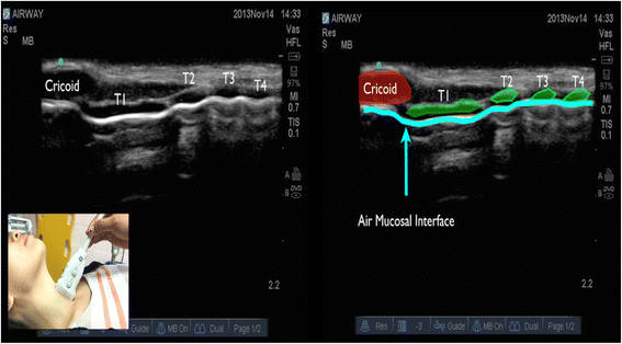 Role Of Upper Airway Ultrasound In Airway Management
 Regional Anesthesia And Ultrasound Central Line Mannequin
 The Radiology Assistant Infrahyoid Neck
 Normal Neck Anatomy And Method Of Performing Ultrasound

 Ultrasonography
 Chapter 4 Ultrasound Of The Neck Thyroid And Parathyroid
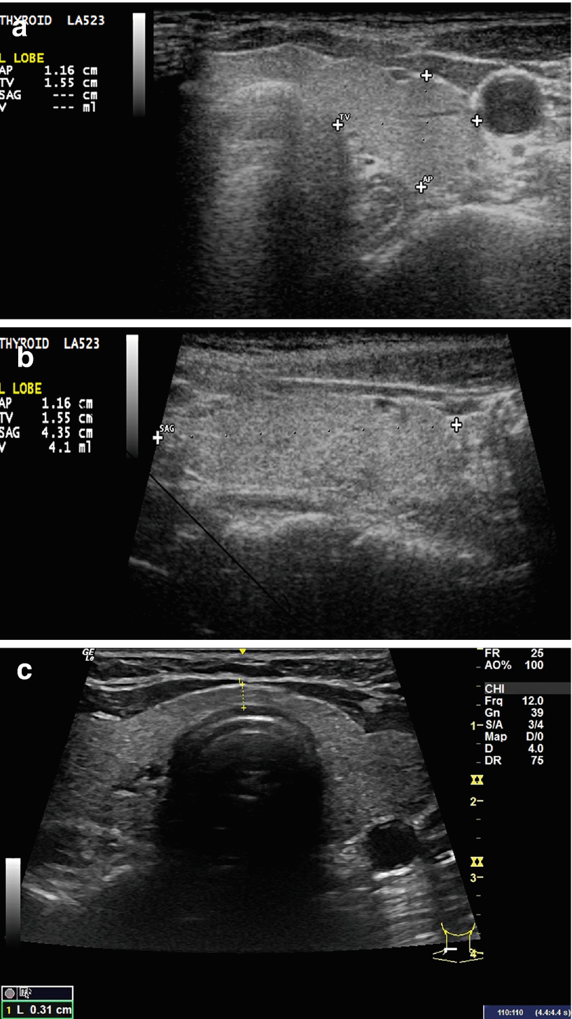
 Normal Thyroid Appearance And Anatomic Landmarks In Neck
 Thyroid Normal Ultrasoundpaedia
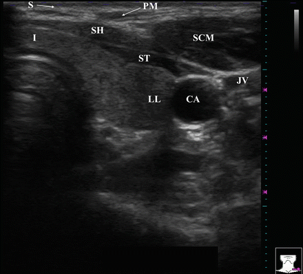
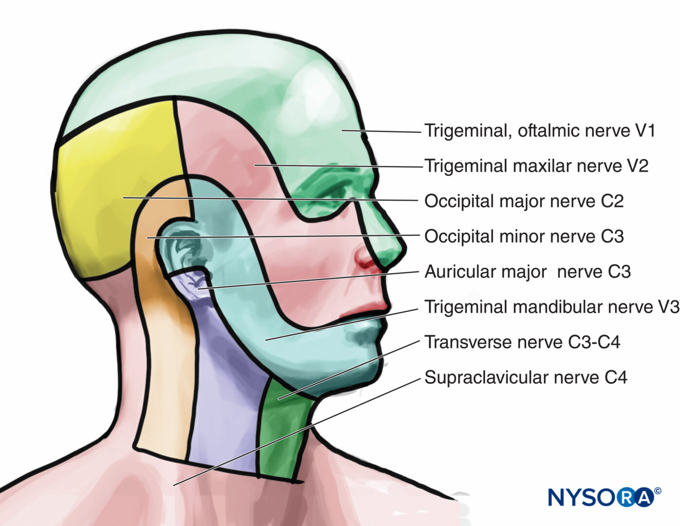 Ultrasound Guided Cervical Plexus Block Nysora
 Ultrasound Of The Pancreas What Normal Looks Like
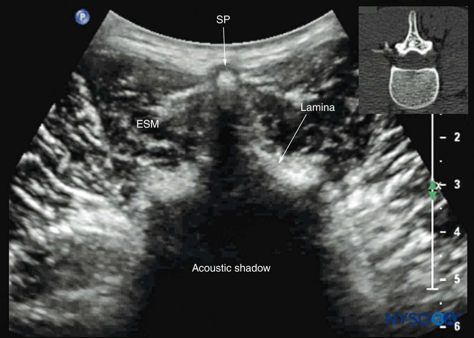 Spinal Sonography And Applications Of Ultrasound For Central
 Normal Thyroid Appearance And Anatomic Landmarks In Neck


Posting Komentar
Posting Komentar