There are three kinds of tmj anatomy pain. Protrusion bilateral contraction of the lateral pterygoid.

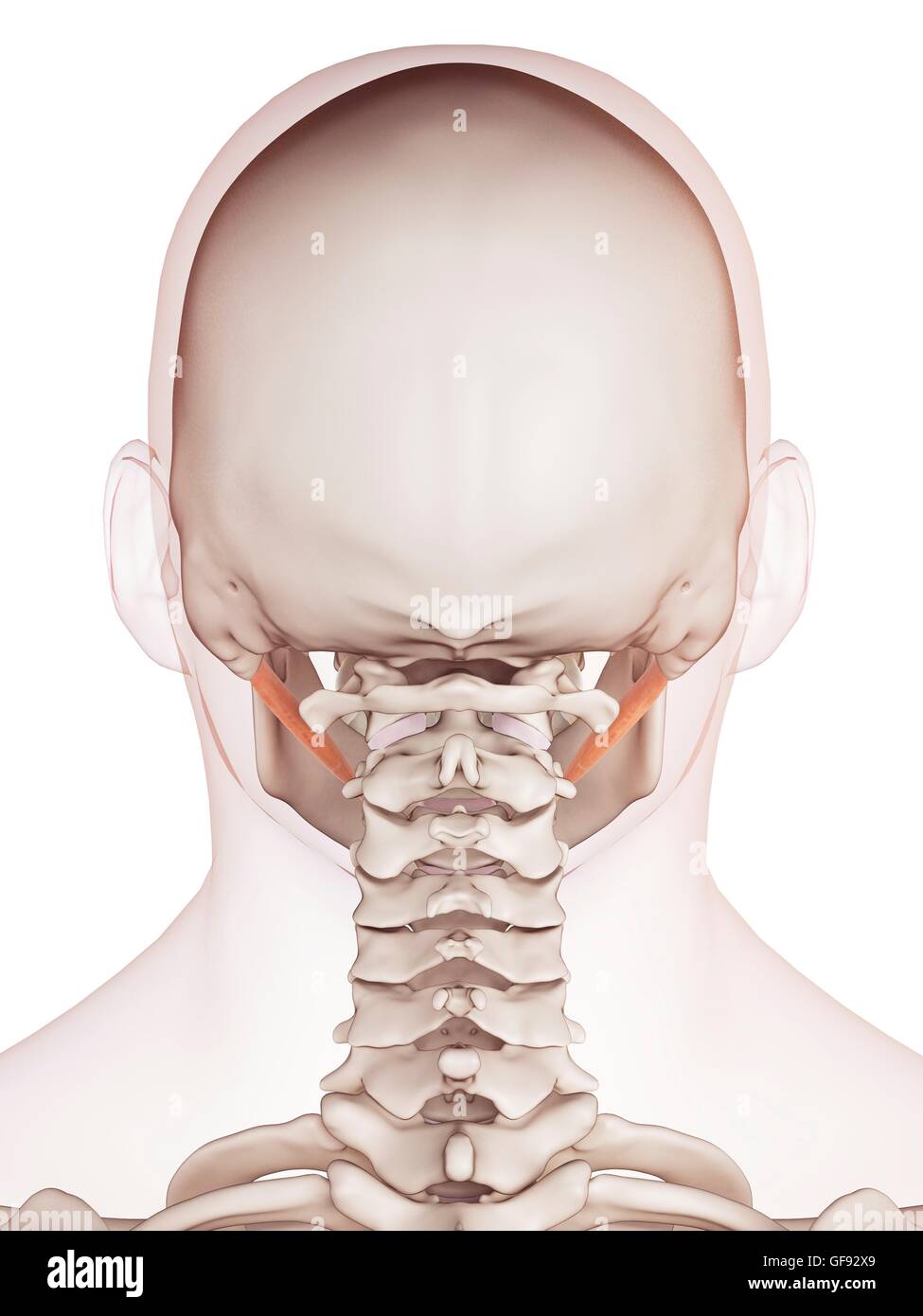
Each of these muscles occurs in pairs with one of each muscle appearing on either side of the skull.
Anatomy jaw muscles. Hypobranchial muscles as the major jaw opening muscle. All four move the jaw laterally. The large muscle which raises the lower jaw and assists in mastication.
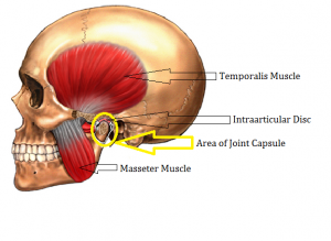
There are four classical muscles of mastication. In regards to jaw anatomy the major joint in the jaw is the temporomandibular joint tmj which connects the lower jaw to the skull temporal bone under the ear. The cardinal mandibular movements of mastication are elevation depression protrusion retraction and side to side movement.
The four main muscles of mastication attach to the rami of the mandible and function to move the jaw mandible. During mastication three muscles of mastication musculi masticatorii are responsible for adduction of the jaw and one the lateral pterygoid helps to abduct it. It is controlled by four bilateral muscles in the face.
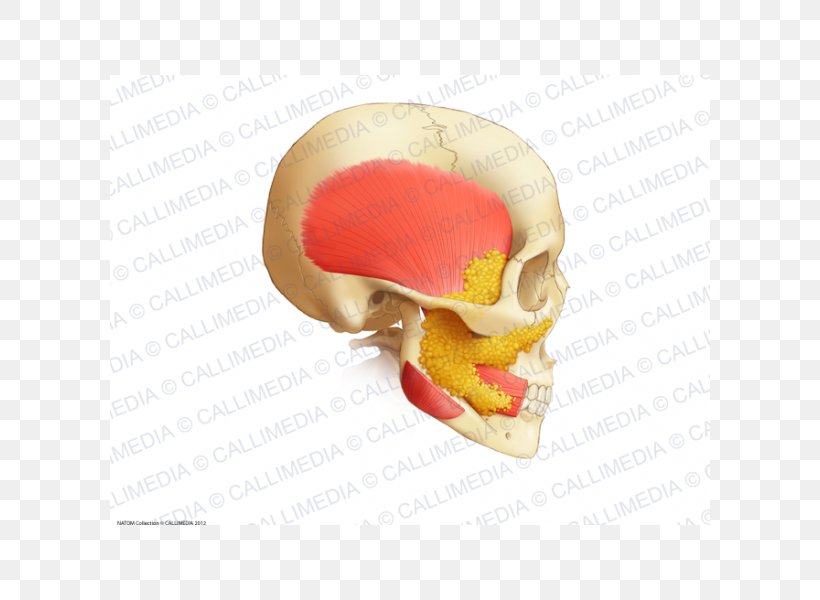
Other muscles usually associated with the hyoid. These muscles including the masseter and temporalis elevate the jaw forcefully during chewing and gently during speech. Some physicians associate disorder in this joint with tiny myofascial trigger points or contractions knots in the overworked or traumatized jaw muscles.
Anatomy of the jaw. These muscles are the masseter the temporalis the medial pterygoid and the lateral pterygoid. Primary muscle discomfort is not really common but overuse as in chewing gum or in south africa biltong in association with disc malfunction can commonly causes jaw facial and sometimes neck pain as well as headache.
Mastication or the act of chewing involves adduction and lateral motion of the jaw bone. There are four muscles the masseter temporalis medial pterygoid and lateral pterygoid. The restructuring of the posterior jaw in mammals leads to the further replacement of this new muscle by the digastric which is a compound muscle made up of parts of the constrictors of the first and second branchial arches.
Retrusion middle and posterior temporalis possibly helped. An extensive complement of tightly interlaced muscles allows the tongue a range of complex movements for chewing and swallowing as well as the important function of producing speech. The muscles of mastication are a group of muscles associated with movements of the jaw.
Closing masseter anterior and middle temporalis medial pterygoid. The muscles work in combination to pivot the lower jaw up and down and to allow movement of the jaw from side to side. Muscles and jaw movement opening inferior head of lateral pterygoid anterior digastric mylohyoid.
Oral Surgery Associates Tmj Disorder

 Acupuncture Treatment For Jaw Pain
![]() Tmj Anatomy Takes You On A Tour Of This Complex Pain Causing
Tmj Anatomy Takes You On A Tour Of This Complex Pain Causing
 Temporalmandibular Joint By Adam Ahmed Chapter 8 Joints
 Tmj Symptoms A Smile Above Vancouver Cosmetic Dentists

 Human Jaw Muscles Anatomy Human Jaw Muscles Anatomy
 Movements Of The Jaw Conscious Movements

 Masseter Muscle Wikipedia
 Human Jaw Muscles Illustration Stock Photo 112681921 Alamy
 Masseter Muscle Jaw Eye Ear Pain Sensitive Teeth The
 Anatomy Ch 11 Mastication And Tongue Muscles Images
 Muscles In Face Lower Jaw And Head Diagram Quizlet
 Canadian Dental Association
 Clenching Grinding Bay Area Tmj And Sleep Center
 Facial Muscles
 Reconstructed Jaw Muscle Anatomy Of Edmontosaurus 1 Left
 Anatomical Teaching Models Plastic Human Pelvic Models
 Neck Cancer Anatomy Headandneckcancerguide Org
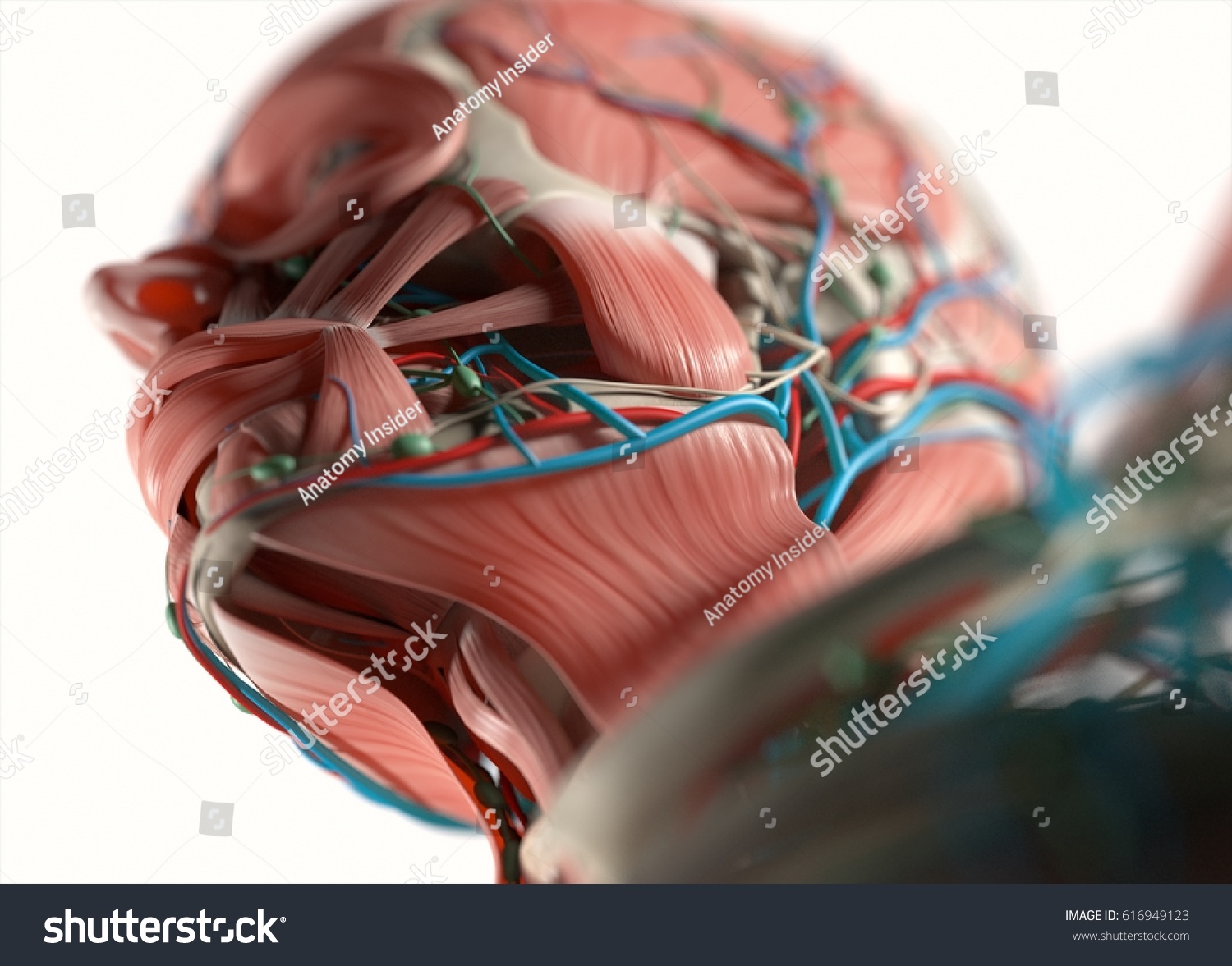 Human Anatomy Face Jaw Nose Muscular Stock Illustration
 Head And Neck Muscles Boundless Anatomy And Physiology
 Massage Therapy For Bruxism Tmj Syndrome
 Medial Pterygoid Muscle An Overview Sciencedirect Topics

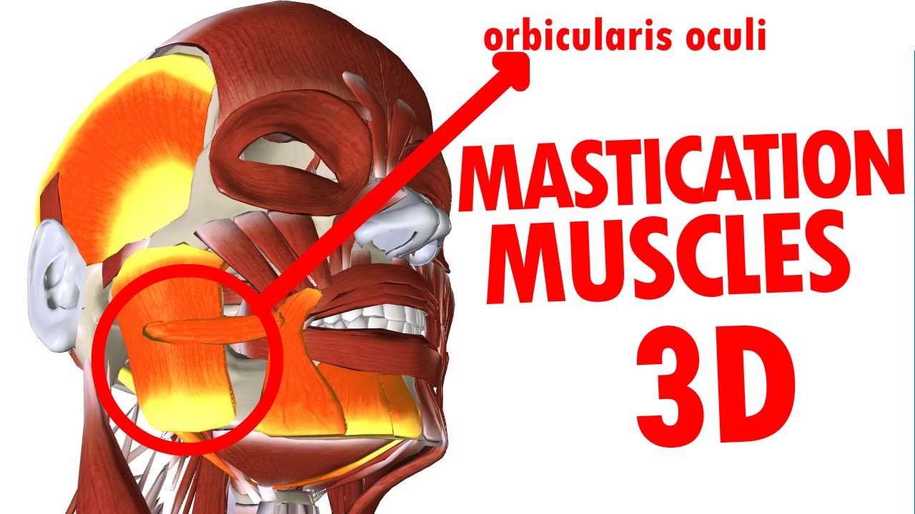 Muscles Of Mastication Jaw And Mandible Face Anatomy Part 3
 Human Anatomy Buccal Fat Pad Jaw Buccinator Muscle Png


Posting Komentar
Posting Komentar