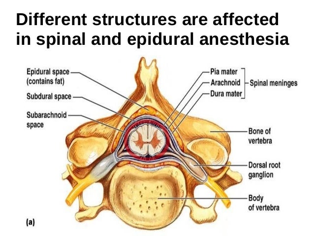
Anatomy of the epidural space vertebral column. The epidural space is an area of spinal anatomy that is located between the vertebral canal and the spinal cord.
 Epidural Anatomy Exhibits
Caudally by sacrococcygeal ligament.

Epidural anatomy. It lies inside the canal but outside the cord. In contact with the inner surface of the dura is another membrane called the arachnoid mater arachnoid. Boundaries cranially by foramen magnum.
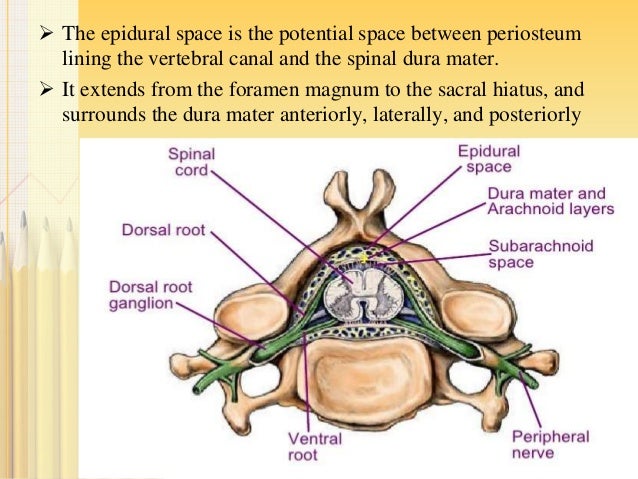
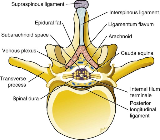
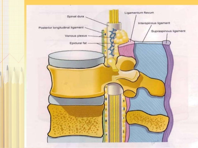
Brain is enclosed within the scull while spinal cord a sausage like structure is enclosed within the spine. The epidural space contains fat epidural veins spinal nerve roots and connective tissue figure 6b the subdural space is a potential space between the dura and the arachnoid and contains a serous fluid. Central nervous system consists of brain and spinal cord.
The cerebrospinal fluid that surrounds the spinal cord is contained by the arachnoid mater. Seven cervical 12 thoracic and five lumbar. Basic anatomy and physiology of central nervous system applicable to epidural and spinal anesthesia is not complicated.
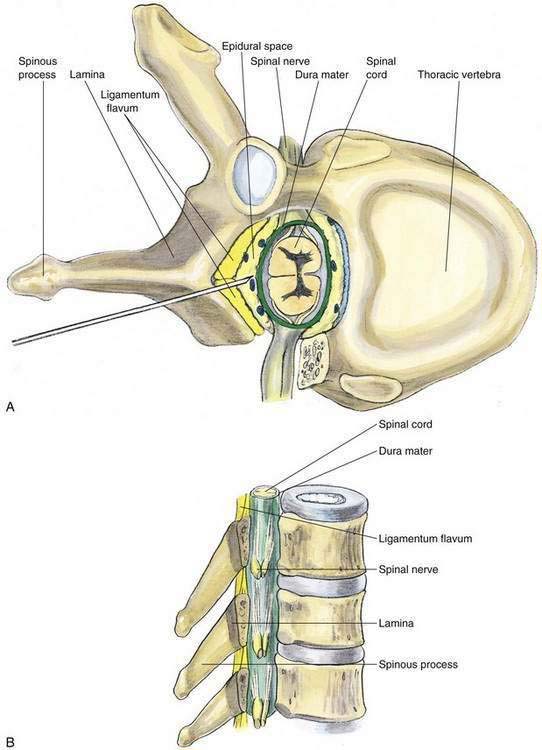
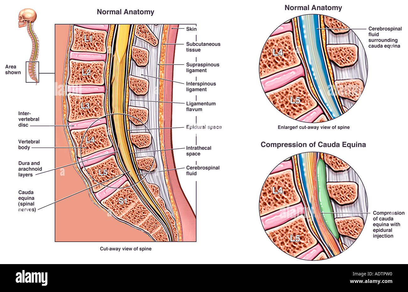
The epidural space is the potential space between periosteum lining the vertebral canal and the spinal dura mater. It extends from the foramen magnum to the sacral hiatus and surrounds the dura mater anteriorly laterally and posteriorly 5. Vertebral anatomy varies according to each level.
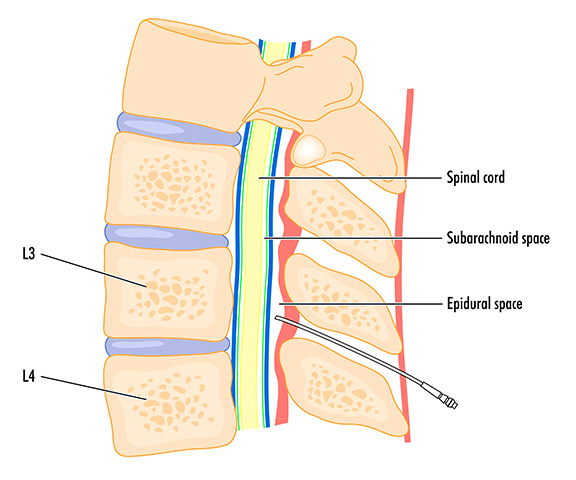
The epidural space is the space inside the bony spinal canal but just outside the dura mater dura. There are 24 individual vertebrae. The five fused sacral vertebrae and the coccyx made up of 35 rudimentary vertebrae are not always classed as being a part of the vertebral column.
The subdural compartment is formed by flat neuroepithelial cells that have long interlacing branches.
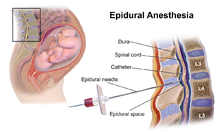
 Spinal Or Epidural Anesthesia
 Anatomy Of Epidural Injection Medical Illustration Medivisuals
 Anatomy Of Spinal Stenosis
 Functional Anatomy Of The Spine For Anesthesia

![]() Spinal Cord Anatomy Structure Tracts And Function Kenhub
Spinal Cord Anatomy Structure Tracts And Function Kenhub

 Epidural Administration Wikipedia
 Neuroanatomy Online Lab 4 External And Internal Anatomy
 Neuraxial Anatomy Nysora
 Anatomy Of The Epidural Needle Soap 50th Annual Meeting
 History Of Epidural Steroid Injections The Burton Report
 Back Clinical Anatomy A Case Study Approach
 Anatomy Of Epidural Space
 Epidural Block Clinical Gate

 Anatomy Of Epidural Space
/epiduralspace-56a05e4f3df78cafdaa149f7.gif) Epidural Space Anatomy And Injections
Epidural Space Anatomy And Injections
Lumbar Facet And Epidural Block Injections Medical
 Epidural Steroid Injection Radiology Key
 Neuraxial Anesthetics And Anatomy Rk Md
In Vivo Images Of The Epidural Space With Two And Three
 Epidural Anatomy Regional Orthopedics Top Physicians
 Spinal Epidural Caudal Blocks Morgan Mikhail S
 Anatomy Epidural Vs Subdural Hematoma Image
 Subarachnoid Hemorrhage Vs Subdural Hematoma Dear Nurses
 Epidural Steroid Injections Pain Management Clinic In
 Anatomy Of Epidural Space
 Epidural Space Stock Photos Epidural Space Stock Images


Posting Komentar
Posting Komentar