The membranous labyrinth is an interconnected group of fluid filled membranous sacs. There are three parts to the ear.
 6 Easy Dog Ear Cleaning Tips You Should Try
Pinched ears usually happen when a dog is very stressed or fearful.

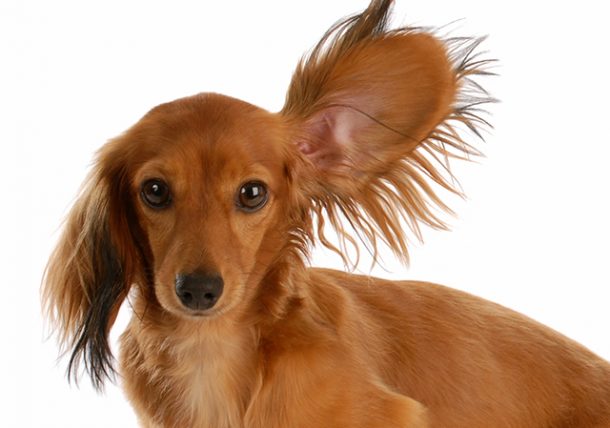
Dog ears anatomy. They can be long short curly or flat. It is composed of a cartilage core and skin. Beyond the ear flap is the ear canal and the eardrum.
The outer ear cannel is separated from the middle ear by a thin membrane called the eardrum or tympanic membrane. In some dogs it is of the floppy variety and in others it is straight or upright. Dog tail anatomy the tail of a dog serves many functions such as non verbal communication and as a rudder in water.
Both species also have ossicles or little bones in the inner ear that vibrate and send signals along the auditory nerve to the brain. The tail isnt just something which wags to show you theyre happy it serves a much bigger function. The inner ear contains the membranous labyrinth which is surrounded by the bony labyrinth.
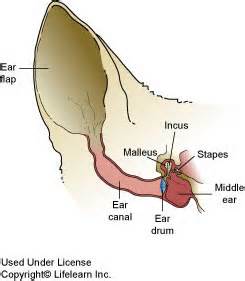
When sound waves hit these layers and the inner ear which is the major functional part of a dogs ear the brain assesses and coordinates the identification of the sound wave. The middle ear includes the eardrum and a small air filled chamber that contains 3 tiny bones. Ear anatomy anatomy of the normal dog ear.
The hammer anvil and stirrup. The anatomy of the middle and inner ear is relatively the same in humans and dogs. It also includes 2 muscles the oval window and the eustachian tube a small tube that connects the middle ear with the back of the nose allowing air to enter.
The ear is turned parallel to the side of the face not back or forward and pulled to the head. The fluid is endolymph. The outer ear middle ear and inner ear.
The anatomy of a dogs ears. Both have an eardrum or tympanic membrane. Ear structure and function in dogs.
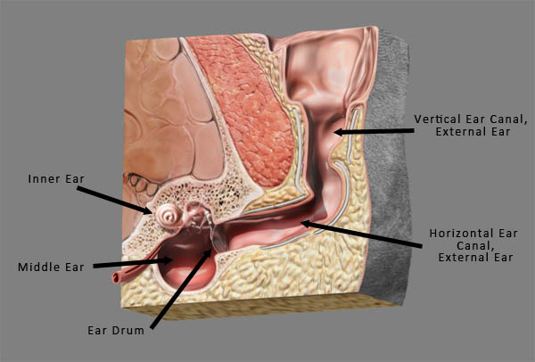
The tail is an extension of the spine so any injuries to the tail can be quite serious. Pinched ears are only visible in floppy eared dogs like goldens labs or hounds. Unlike humans that have a very short ear canal dogs have a long narrow ear canal that makes almost a 90 degree bend as it travels to the deeper parts of the ear.
This is the external most visual portion of the canine ear. The inner ear is located within the petrous temporal bone. The ear flap is part of the outer ear and stands up tall in some dog breeds or flops over in others.
Moreover associated anatomical bodies such as cartilages nerves vascular supply glands and the ear cavity assists in the normal functioning of ear and helps the body to maintain balance.
 Dog Ear Canal Diagram Dog Anatomy Ear Canal Diagram Anatomy
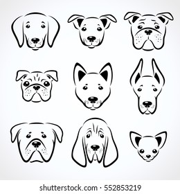 Dog Ears Images Stock Photos Vectors Shutterstock
 Cleaning Your Dogs Ears Canine Ear Diagram Ear Cleaning
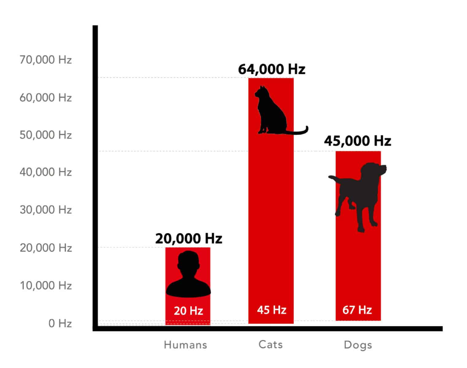 Hearing Frequencies Anatomy Human Cat Dog Icalmpet
 Ear Structure And Function In Dogs Dog Owners Merck
 Ear Anatomy Physiology Wikivet English
Vet Tips To Recognize And Treat Pet Ear Problems The
Anatomical Studies Of Canine Vascular And Ligamentous Ear
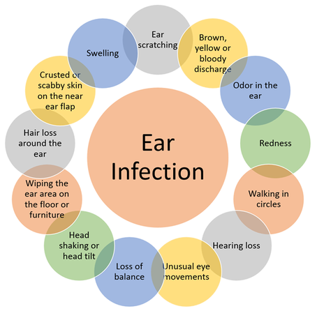
 Dog Ear Infections Learn How To Treat Prevent Your Vet

 6 Proven Steps For Healthy Dog Ears How To Clean Dog Ears

 5 Best Dog Ear Cleaners W Free Dog Ear Cleaning Guide

 What Is A Henry S Pocket And Why Does My Dog Have One
 What S With That Slit On Your Cat S Ear And More On Feline
 Will It Hurt A Dog S Ears To Be Around The Sound Of Gunfire
 How To Keep Your Dog S Ears Clean Canine Cleanliness Tips
 What S With That Slit On Your Cat S Ear And More On Feline
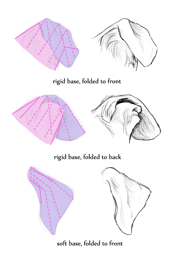 How To Draw Animals Dogs And Wolves And Their Anatomy
 Overview Of Otitis Media And Interna Ear Disorders Merck
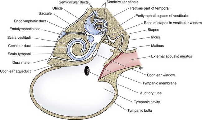 Middle And Inner Ear Veterian Key
 Dog Ear Anatomy Ear Anatomy Cleaning Dogs Ears Dog Anatomy

 Four Steps To Keep Your Dog S Ears Healthy By Lindy Callahan


Posting Komentar
Posting Komentar