Its strength and joint function facilitate running jumping walking up stairs and raising the body onto the toes. These tendons help your extensor muscles pull your foot upwards which is necessary for walking14.
 Ankle Foot Anatomy
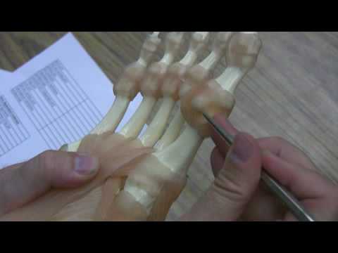
Tendons allow movements by connecting the muscles to bones.

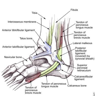
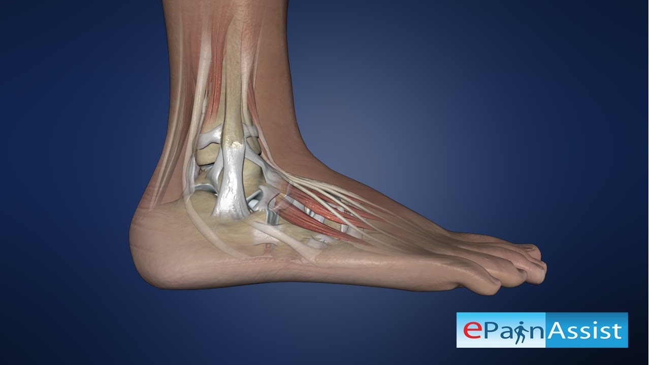
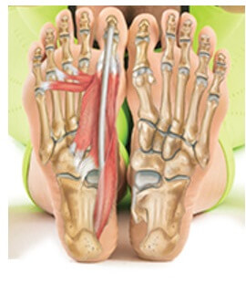
Anatomy of the foot tendons and ligaments. Foot tendons and ligaments diagram 9 photos of the foot tendons and ligaments diagram foot anatomy diagram foot joint diagram foot sprain diagram foot tendons and ligaments pain leg tendon diagram peroneal tendonitis foot foot anatomy diagram foot joint diagram foot sprain diagram foot tendons and ligaments pain leg tendon diagram. In humans the foot is one of the most complex structures in the body. Muscles tendons and ligaments run along the surfaces of the feet allowing the complex movements needed for motion and balance.
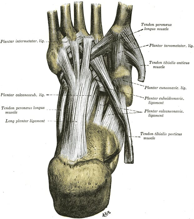
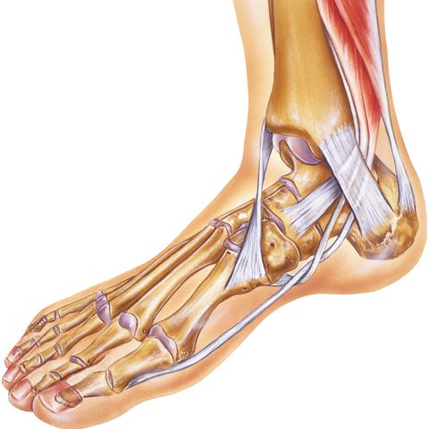
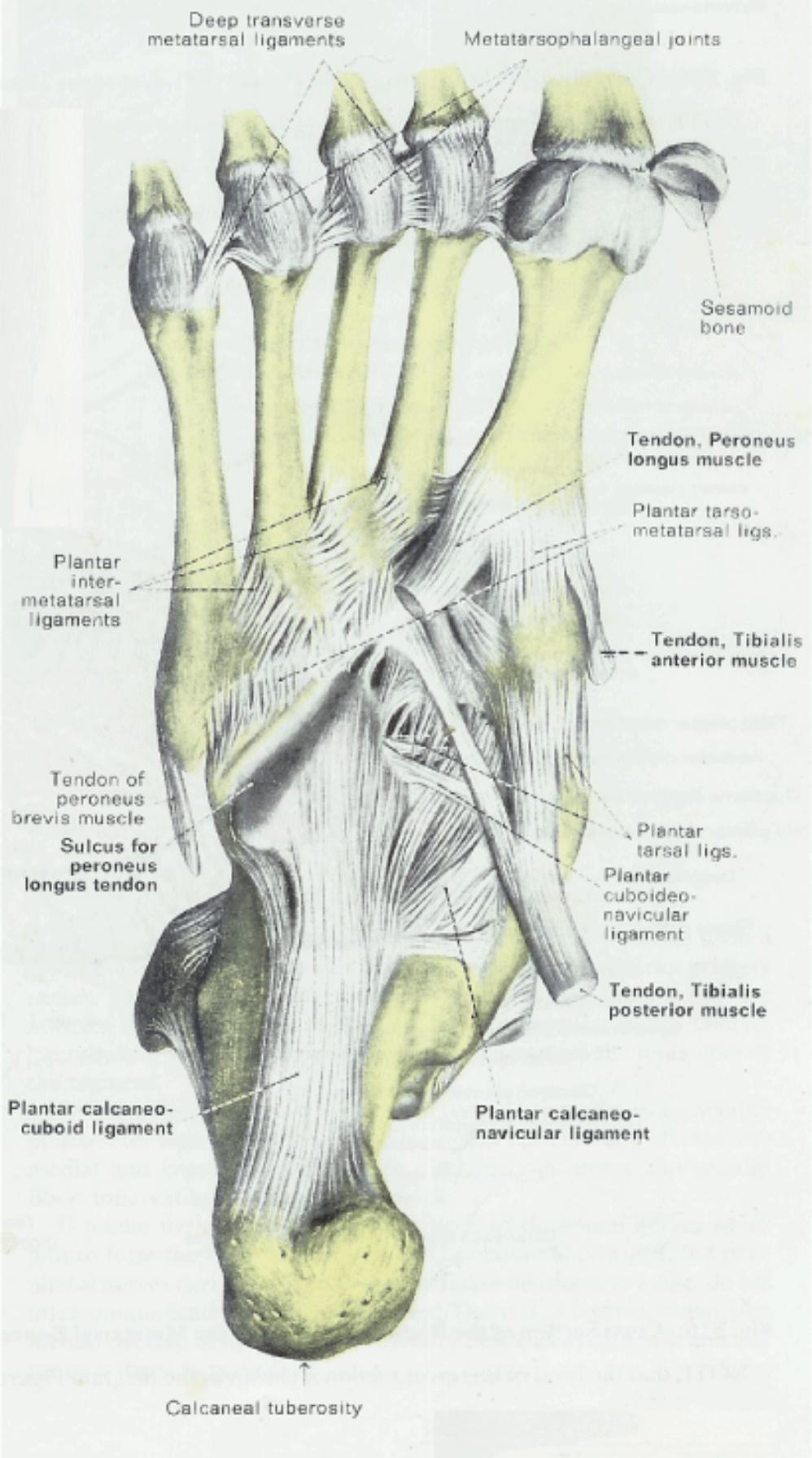
Ligaments hold the tendons in place and stabilize the joints. The ligament which runs along the sole of the foot from the heel to the toes forms the arch. Due to less blood flow in ligaments sprains are not easily recovered and long term damage results on the ligaments.
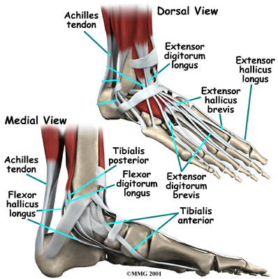
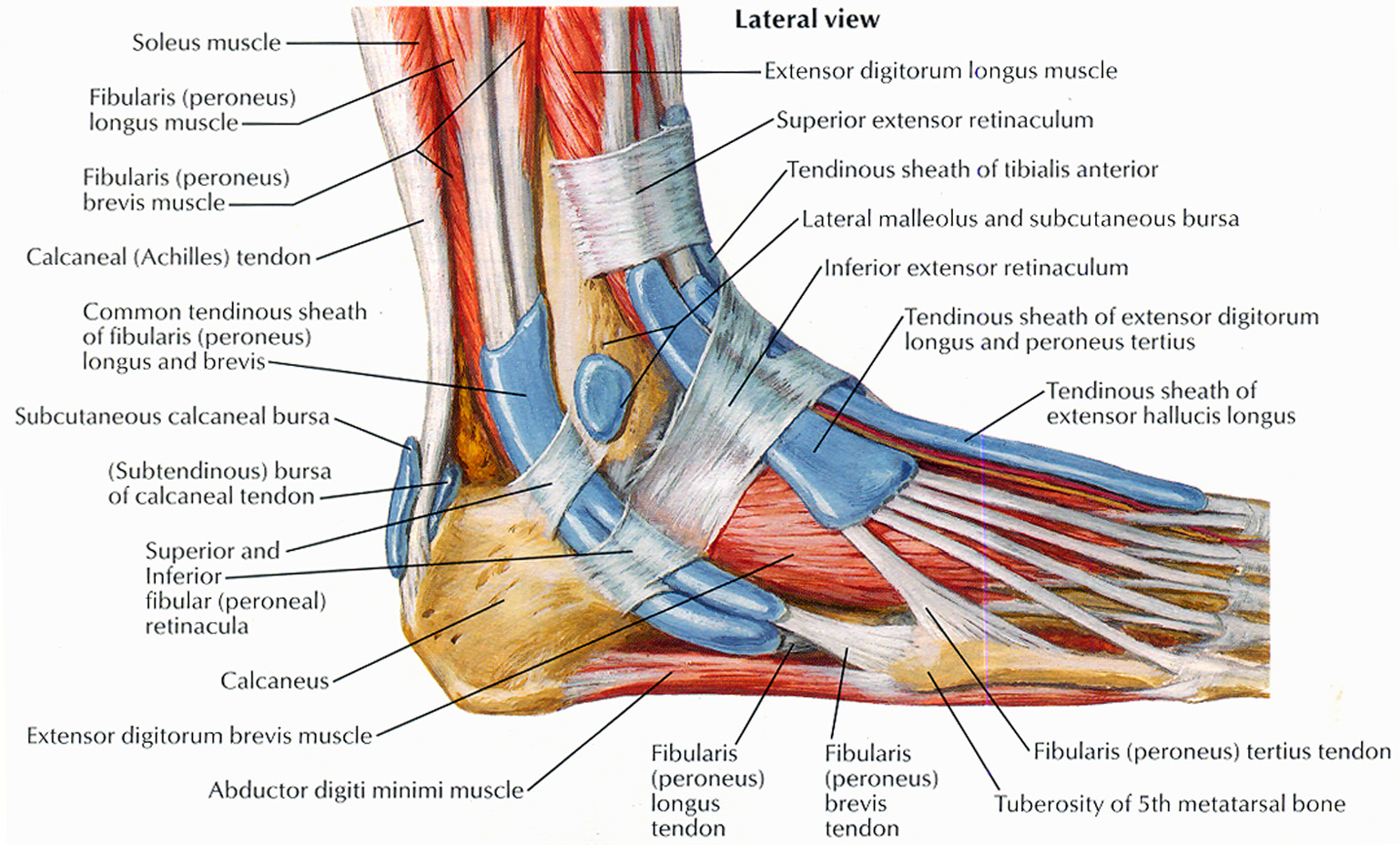
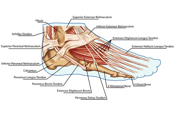
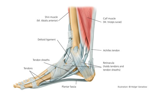
Extensor tendinitis happens when the tendons on top of your foot become inflamed. The largest and strongest tendon of the foot is the achilles tendon which extends from the calf muscle to the heel. Plantar fascia the longest ligament of the foot.
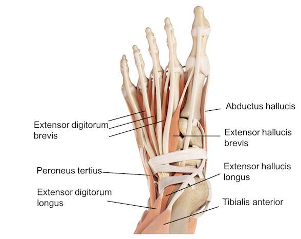
The function of ligaments is to attach bones to bones and help to stabilize them to one another. The two main extensor foot tendons are the extensor hallucis longus and the extensor digitorum longus. It is made up of over 100 moving parts bones muscles tendons and ligaments designed to allow the foot to balance the bodys weight on just two legs and support such diverse actions as running jumping climbing and walking.
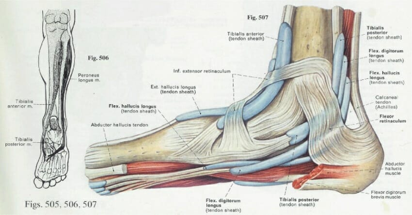
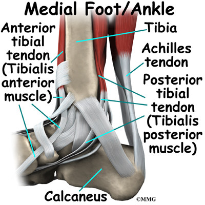
Foot ankle anatomy muscles tendons and ligaments. By stretching and contracting the plantar fascia helps us balance and gives the foot strength for walking. The calcaneus heel bone is the largest bone in the foot.
Foot anatomy diagram foot joint diagram foot sprain diagram foot tendons and ligaments pain leg tendon diagram peroneal tendonitis foot foot anatomy diagram foot joint diagram foot sprain diagram foot tendons and ligaments pain leg tendon diagram peroneal tendonitis. The thick bands of tissues that connect muscles to bones are called tendons. Medial ligaments of the foot arch side of the foot ligaments are strong dense and flexible bands of fibrous connective tissue.
The main ligaments of the foot are.
Ankle Anatomy
 Foot Anatomy Bones Ligaments Muscles Tendons Arches

 An Overview Of The Inferior Check Ligament In Horses
 Human Anatomy Ligaments And Tendons Of The Foot
 Foot Ankle Anatomy Pictures Function Treatment Sprain Pain
 Ligament Injury Tendon Injury What S The Difference
 Ligaments Of The Foot Muscles Tendons Ligaments Of The
 Peroneal Tendon Syndromes Practice Essentials Epidemiology
 Ligaments Of The Foot Tendons In The Foot Wedding Love
 Achilles Tendon Anatomy And Importance
![]() Tendon Sheaths In The Foot Anatomy Kenhub
Tendon Sheaths In The Foot Anatomy Kenhub
 Ligaments And Tendons Of The Foot
Arthritis Of The Foot And Ankle Orthoinfo Aaos
 Foot Anatomy Bones Ligaments Muscles Tendons Arches
 Common Running Ankle Injuries Everything You Need To Know
 Arches Of The Foot Physiopedia
 Ankle Anatomy Eorthopod Com
 Ankle Joint Anatomy Explained Bones Joints Ligaments
 Figure Interior Ligaments Of The Left Statpearls
 Plantar Fasciitis And Foot Pain In Nursing
 Novobrace Tendonitis Desmitis And Soft Tissue Injury
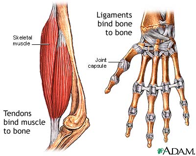 Tendon Vs Ligament Medlineplus Medical Encyclopedia Image
 Ligament Vs Tendon What S The Difference
 Foot And Ankle Anatomy Bones Muscles Ligaments Tendons
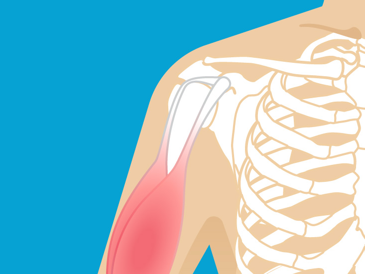
 Ligaments Muscles And Tendons
 The Arches Of The Foot Longitudinal Transverse


Posting Komentar
Posting Komentar