The ear the eye the nose and sinuses the salivary glands and the oral cavity. The cerebellum is responsible for coordination and balance.
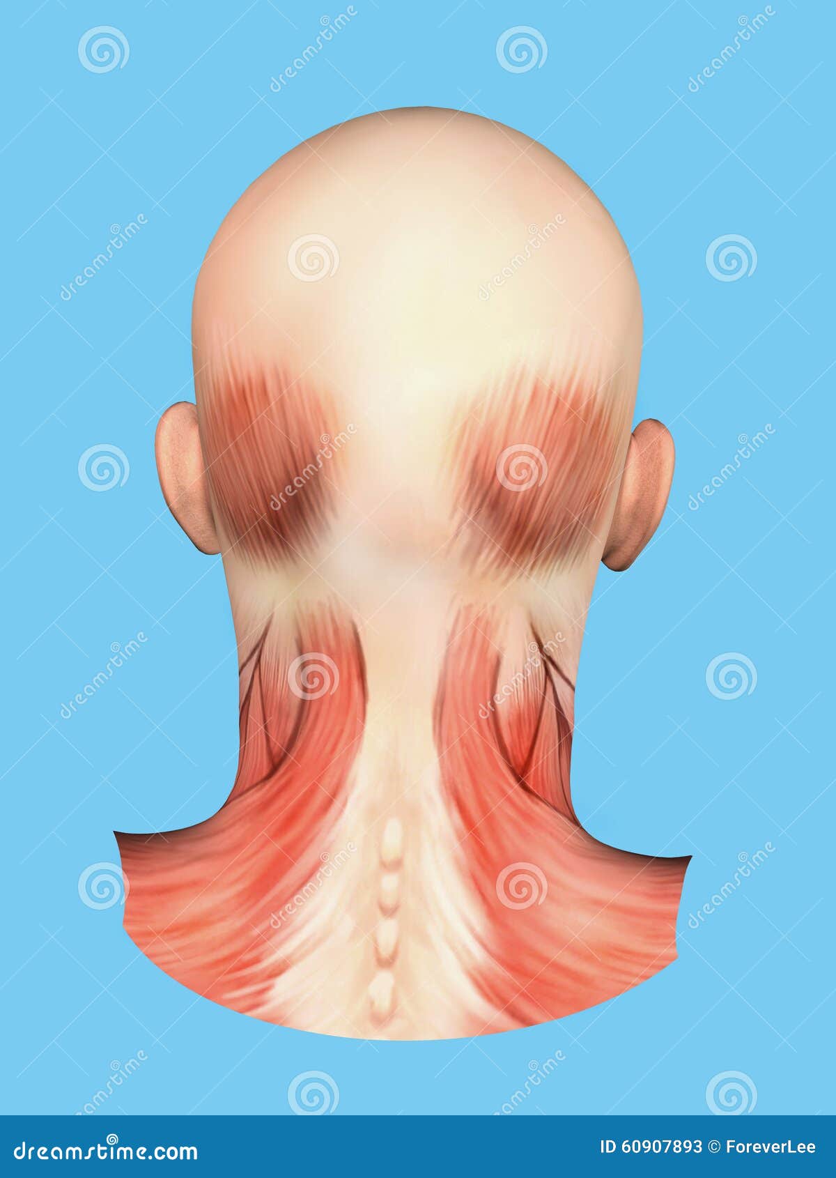 Anatomy Of Muscles On Back Of Head Stock Illustration
The external ear the middle ear and the inner ear.

Back of the head anatomy. Their afferent vessels drain the occipital region of the scalp while their efferents pass to the superior deep cervical glands. Superficial dissections of the head and neck as seen in the gallery show the many different muscles that are required for movement plus those that control facial expression. The skeletal section of the head and neck forms the top part of the axial skeleton and is made up of the skull hyoid bone auditory ossicles and cervical spine.
We use cookies to ensure that we give you the best experience on our website. Working in pairs on the left and right sides of the body these muscles control the flexion and extension of the head and neck. Tonsils are located in the back of throat and are part of the lymphatic system.
A retracted head is seen in acute meningitis cerebral abscess tumor thrombosis of the superior longitudinal sinus acute encephalitis laryngeal obstruction tetanus hydrophobia epilepsy spasmodic torticollis strychnine poisoning hysteria rachitic conditions and painful neck lesions at the back. The occipital glands lymphoglandulæ occipitales one to three in nu ber are placed on the back of the head close to the margin of the trapezius and resting on the insertion of the semispinalis capitis. The lymphatics of the head face and neck.
Tonsillitis is a fairly common infection of the. The brain is also divided into several lobes. The organs of the head include.
Teachme anatomy part of the teachme series the medical information on this site is provided as an information resource only and is not to be used or relied on for any diagnostic or treatment purposes. The external ear functions to capture and direct sound waves through the external acoustic meatus to reach the tympanic membrane ear drum. If you continue to use this site we will assume that you are happy with it.
They catch and kill germs that enter the body through the mouth. The cerebellum is at the base and the back of the brain. The muscle anatomy of the head and neck is a fascinating area with the the neck also containing the 7 vertebrae of the part of the spine called the cervical curve.
The ear can be divided in to three sections. They move the head in every direction pulling the skull and jaw towards the shoulders spine and scapula. The head rests on the top part of the vertebral column with the skull joining at c1 the first cervical vertebra known as the atlas.
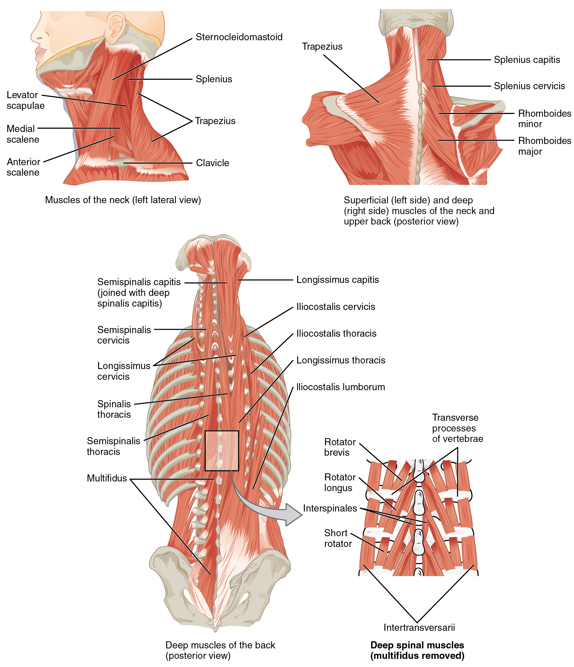 11 3 Axial Muscles Of The Head Neck And Back Anatomy And
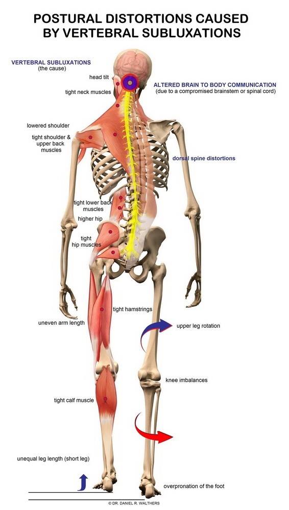 Get Neck And Lower Back Pain Relief San Antonio Thrive
 Picture Of Human Anatomy Backside Stock Photos Page 1
 Phace Syndrome Handbook Abnormalities Of The Head And Neck
 Back Human Skull View From Behind
:watermark(/images/watermark_only.png,0,0,0):watermark(/images/logo_url.png,-10,-10,0):format(jpeg)/images/anatomy_term/trapezius-muscle-3/sx5vl2FteBqX9H04JXGUvQ_Musculus_trapezius_2.png) Back Muscles Anatomy And Functions Kenhub
Back Muscles Anatomy And Functions Kenhub
The Skull Anatomy And Physiology Openstax
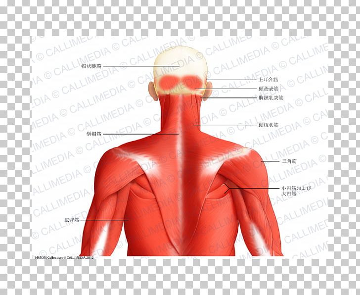 Muscle Posterior Triangle Of The Neck Head And Neck Anatomy
 Human Head Neck Skull Anatomy Medical Anatomical Chart
Headache Symptoms Causes Diagnosis Headache Treatment
 Complete Anatomy Complete Anatomy
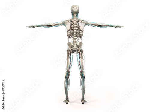 Human Anatomy Showing Back Full Body Head Shoulders And
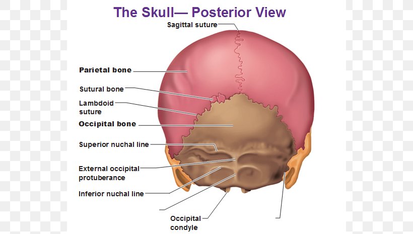 Human Body Skull Anatomy External Occipital Protuberance
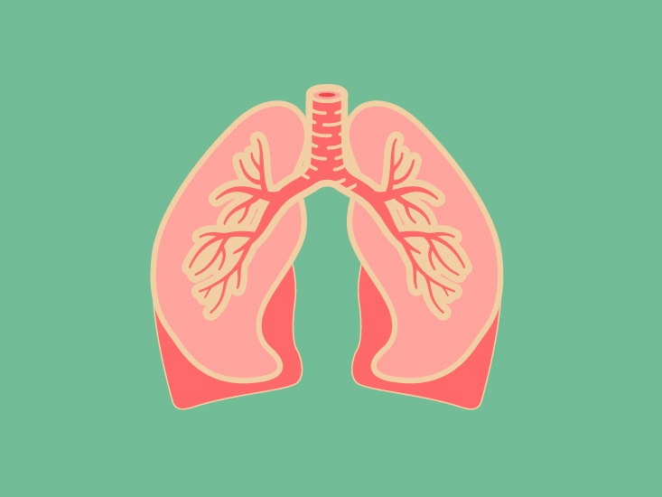 Sphenoid Sinus Anatomy Diagram Location Body Maps
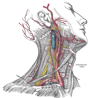 Head And Neck Anatomy Wikipedia
The Skull Anatomy And Physiology Openstax
 Massage Therapy For Tension Headaches
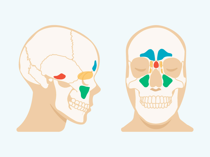 Sinus Cavities In The Head Anatomy Diagram Pictures
 Female Head Muscles Anatomy Back Clipart K20223320
 4 Views Of The Head Back And Spine From An Atlas Of Human
 Human Back Human Head Human Body Muscle Png 900x550px
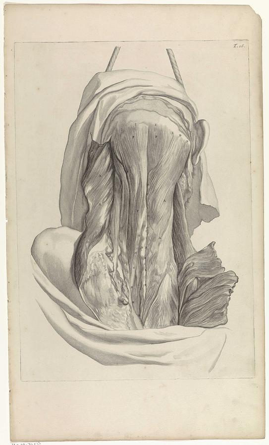 Anatomical Study Of The Back Of The Head Pieter Van Gunst After Gerard De Lairesse 1685
 Figure Anatomy Of The Brain The Pdq Cancer
 Anatomical Chart Human Skull Sticky Back

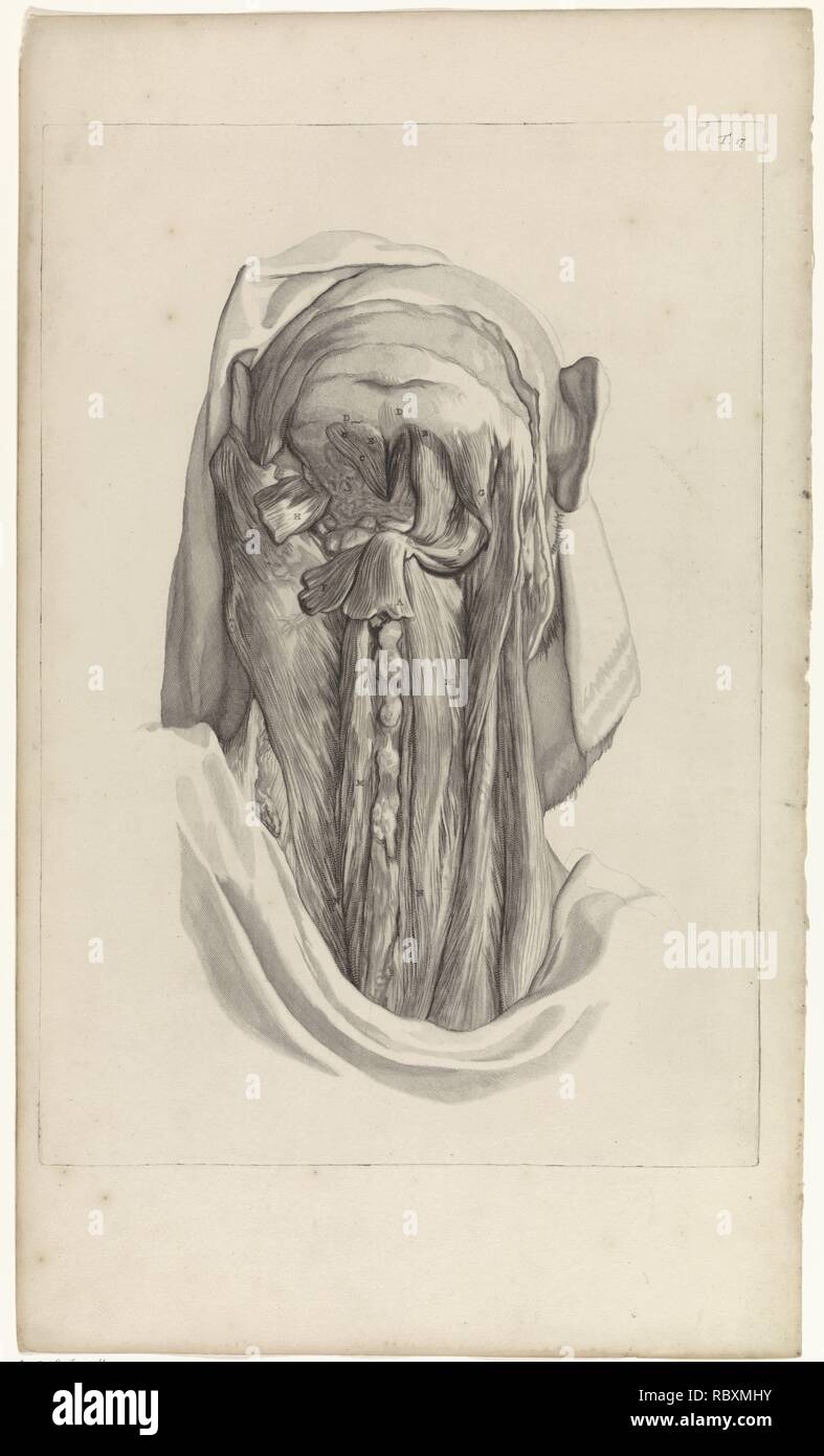 Anatomical Study Of The Muscles Of The Back Of The Head And
 Muscles Advanced Anatomy 2nd Ed



Posting Komentar
Posting Komentar