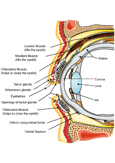
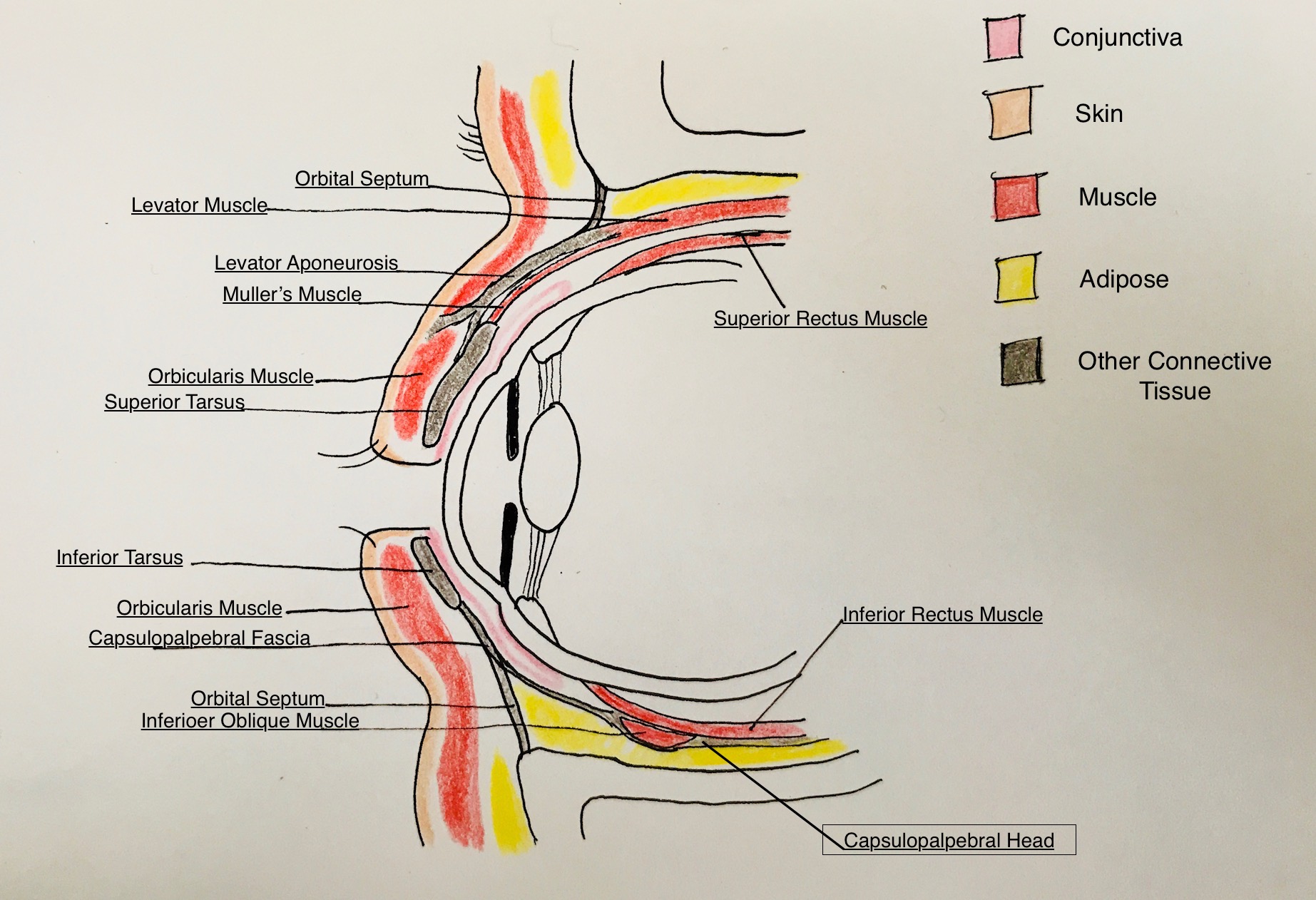
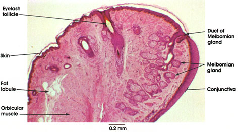
Eyelid and orbit anatomy anatomy of the eyelid. The eyelid is primarily made of skin.
 The Cyclopaedia Of Anatomy And Physiology Anatomy
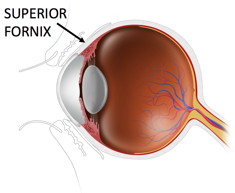
The orbit is the bony eye socket of the skull.

Inner eyelid anatomy. Overview of external anatomy. Vision is by far the most used of the five senses and is one. Meibomian gland openings reside here and exude the lipid layer of the tear film.
The eyelid margin is a specialized location between the skin and conjunctiva. Eyelid movable tissue consisting primarily of skin and muscle that shields and protects the eyeball from mechanical injury and helps to provide the moist chamber essential for the normal functioning of the conjunctiva and cornea. The eyelids serve to protect the eye from foreign matter such as dust dirt.
The lateral canthal angle is 2 mm higher than the medial canthal angle in europeans. The eyelids have haired skin on the outside layer and are lined on the inside with conjunctiva. Skin and subcutaneous tissue.
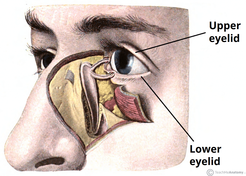
The conjunctiva is the mucous membrane that lines the eyelid and covers the visible portion. The upper eyelid starts at the eye and extends up words joined the skin. The description above only offers a superficial overview of the anatomy.
An epicanthic fold the skin fold of the upper eyelid covering the inner corner medial canthus of the eye may be present based on various factors including ancestry age and certain medical conditions. It is 3 mm higher in asians. And is inserted on the peripheral margins of the tarsal plate.
The lower lid rests at the inferior limbus and peaks 1 mm lateral to the center of the pupil. The upper lid naturally rests 1 to 2 mm below the superior limbus and peaks 1 mm medial to the center of the pupil. Anatomy and physiology of the eye eye anatomy facts.
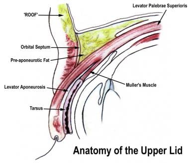
The anterior lamella is comprised of the skin and orbicularis oculi muscle and the posterior lamella is made up of the tarsal plate and conjunctiva. The lower eyelid margin rests at the level of the lower limbus. In youths the upper lid margin rests at the upper limbus while in adults it rests 15 mm below the limbus.
In some populations the trait is almost universal specifically in east asians and southeast asians where a majority up to 90 in some estimations of adults have this feature. In the upper lid it arises from the fibres of lps muscle and in the lower lid from prolongation of the inferior rectus muscle. The anterior and posterior lamellae.
The gray line is considered the junction of the anterior and posterior lamellae. The inferior eyelid fold inferior palpebral sulcus. The upper and lower eyelids along with the upper and lower puncta oppose the globe.
The upper eyelid skin crease superior palpebral sulcus is approximately 811 mm superior to the eyelid margin. Layer of non striated muscle fibres. The eyelid is structurally divided into two anatomical lamellae.
It consist of the palpebral muscle of muller which lies deep to the septum orbitale in both the lids. It is formed by the attachment of the superficial insertion of levator aponeurotic fibers 89 mm in men and 911 mm in women.
 Blepharitis
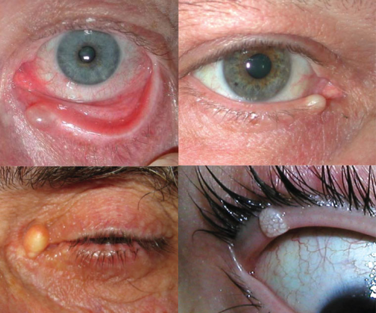
 Pictures Of Styes And Chalazia In Your Eye
 Blepharoplasty Eye Lift Eyelid Surgery
 Pictures Of Styes And Chalazia In Your Eye
 Eye Anatomy Glaucoma Research Foundation
 Anatomy Of The Eyelids Springerlink
 Anatomy Of Lower Eyelid Of Humans Human Eye Anatomy Parts Of
 Eyelid Muscle An Overview Sciencedirect Topics
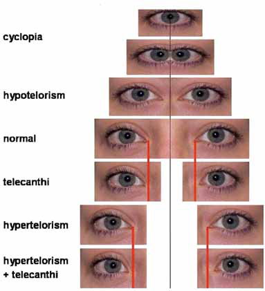 Elements Of Morphology Human Malformation Terminology
 Eyelid Anatomy Overview Surface Anatomy Skin And
 Vision And The Eye S Anatomy Healthengine Blog
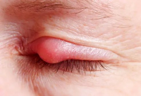
 Chalazion
 Racgp Eyelid Lesions In General Practice
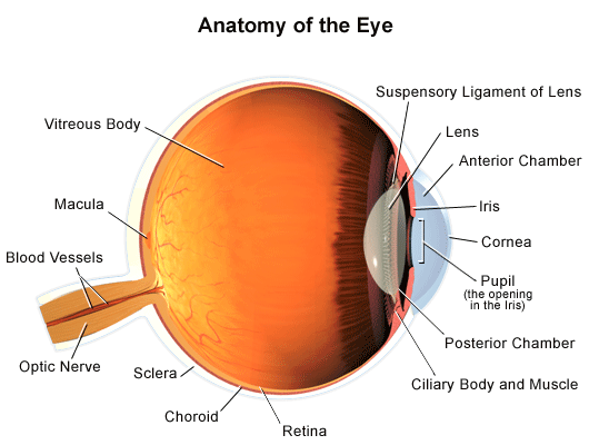
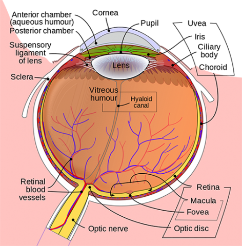 Anatomy Of The Eye Kellogg Eye Center Michigan Medicine
 Aurolab Anatomy

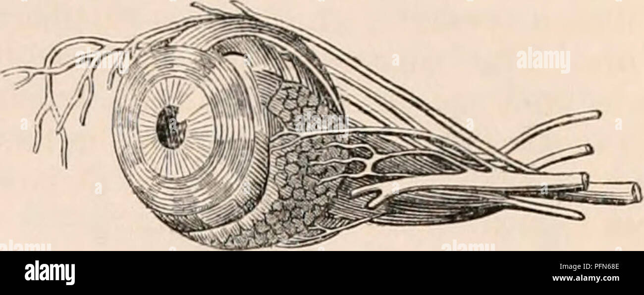
 Eye Structure And Function In Dogs Dog Owners Merck

 Anatomy Of The Eye
Conjunctivitis In Dogs Vca Animal Hospital
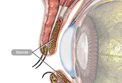
 The Eyelids Conjunctiva Muscles Lacrimal Glands
 Third Eyelid Gland Prolapse In Dogs Mspca Angell
 Sty Stye Causes Treatment Symptoms Remedies Pictures
 Eye Structure And Function In Dogs Dog Owners Merck
 Anatomy Atlases Atlas Of Microscopic Anatomy Section 1 Cells


Posting Komentar
Posting Komentar