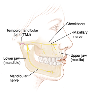
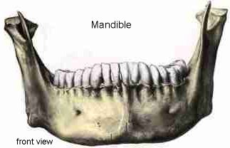
It forms the lower jaw and acts as a receptacle for the lower teeth. The mandible articulates with the neurocranium at the temporomandibular joints tmjs.
![]() Jaw Pain If It Is A Tmj Problem Physiotherapy Can Help
Jaw Pain If It Is A Tmj Problem Physiotherapy Can Help
It forms the lower jaw and holds the lower teeth in place.

Jaw anatomy. The mandible is a u shaped lower jawbone and the largest strongest bone in the face figures 1 and 2 and the only one that can move significantly. Four different muscles connect. Movement of the lower jaw opens and closes the mouth and also allows for the chewing of food.
Definition csp bony structure of the mouth that holds the teeth. It also articulates on either side with the temporal bone forming the temporomandibular joint. Consists of the mandible and the maxilla.
Definition msh bony structure of the mouth that holds the teeth. The mandible lower jaw or jawbone is the largest strongest and lowest bone in the human face. The lower set of teeth in the mouth is rooted in the lower jaw.
Jaw either of a pair of bones that form the framework of the mouth of vertebrate animals usually containing teeth and including a movable lower jaw and fixed upper jaw maxilla. It is the only movable bone of the skull. Jaw c0022359 definition nci the bones of the skull that frame the mouth and serve to open it.
The mandible or lower jaw is the bone that forms the lower part of the skull and along with the maxilla upper jaw forms the mouth structure. Mandible supports the lower teeth and provides attachment for muscles of mastication and facial expression. The mandible sits beneath the maxilla.
Dysfunction of the tmj can cause severe pain and lifestyle limitation. The mandible located inferiorly in the facial skeleton is the largest and strongest bone of the face. Tmj anatomy the temporomandibular joint tmj or jaw joint is a bi arthroidal hinge joint that allows the complex movements necessary for eating swallowing talking and yawning.
The bones that hold the teeth. The bone is formed in the fetus from a fusion of the left and right mandibular prominences and the point where these sides join the mandibular symphysis is still visible as a faint ridge in the midline. It consists of the mandible and the maxilla.
Jaws function by moving in opposition to each other and are used for biting chewing and the handling of food. Like other symphyses in the body this is a midline articu.
 Understanding Orthognathic Anatomy And Problems Medcor
 Human Skull Replica Life Size Articulating Mandible Jaw
 Amazon Com Anatomy Jaw Nervous System Print Sra3 12x18
 The Mandible Lower Jaw Human Anatomy
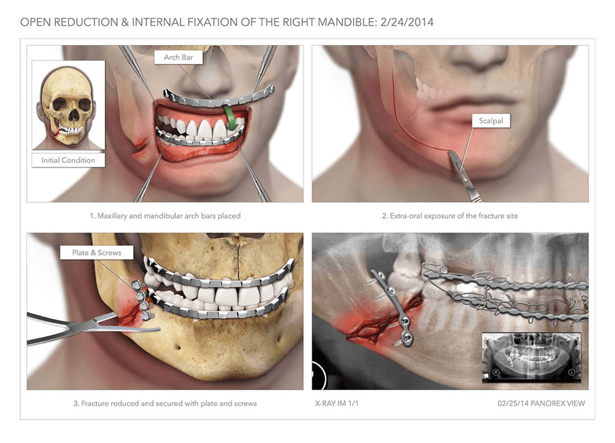 Anatomy Of The Jaw And Teeth High Impact Visual
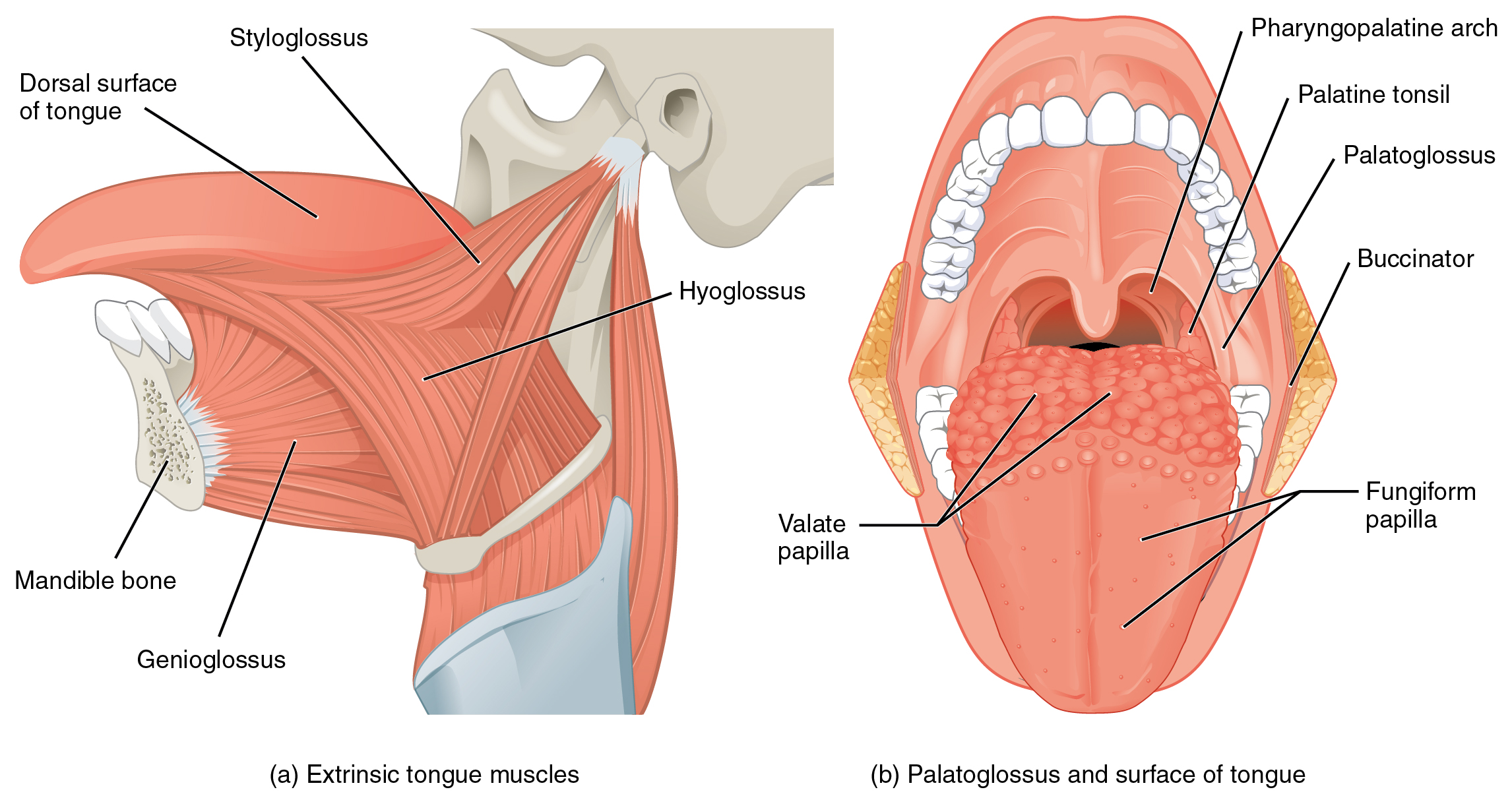 11 3 Axial Muscles Of The Head Neck And Back Anatomy And
 Orthognathic Surgery Wikipedia
 Anatomy Of The Neck And Jaw Anatomy Of The Jaw And Neck
 Muscles Of The Tmj Jaw Pain Headaches Henry S Natural
:background_color(FFFFFF):format(jpeg)/images/library/7639/IkZJJPMujD6IQ2e7r8yiNA_mandible_latin.jpg) The Mandible Anatomy Structures Fractures Kenhub
The Mandible Anatomy Structures Fractures Kenhub
 Real Human Mandibular Jaw Anatomy With Teeth 3d Print Model
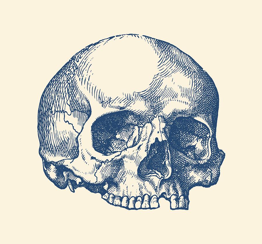 Human Skull No Jaw Simple View
 Mandible Structure Muscular Attachments Anatomy
 Teeth And Jaw Bone Anatomy Print
 Skull Anatomy Pictures And Information
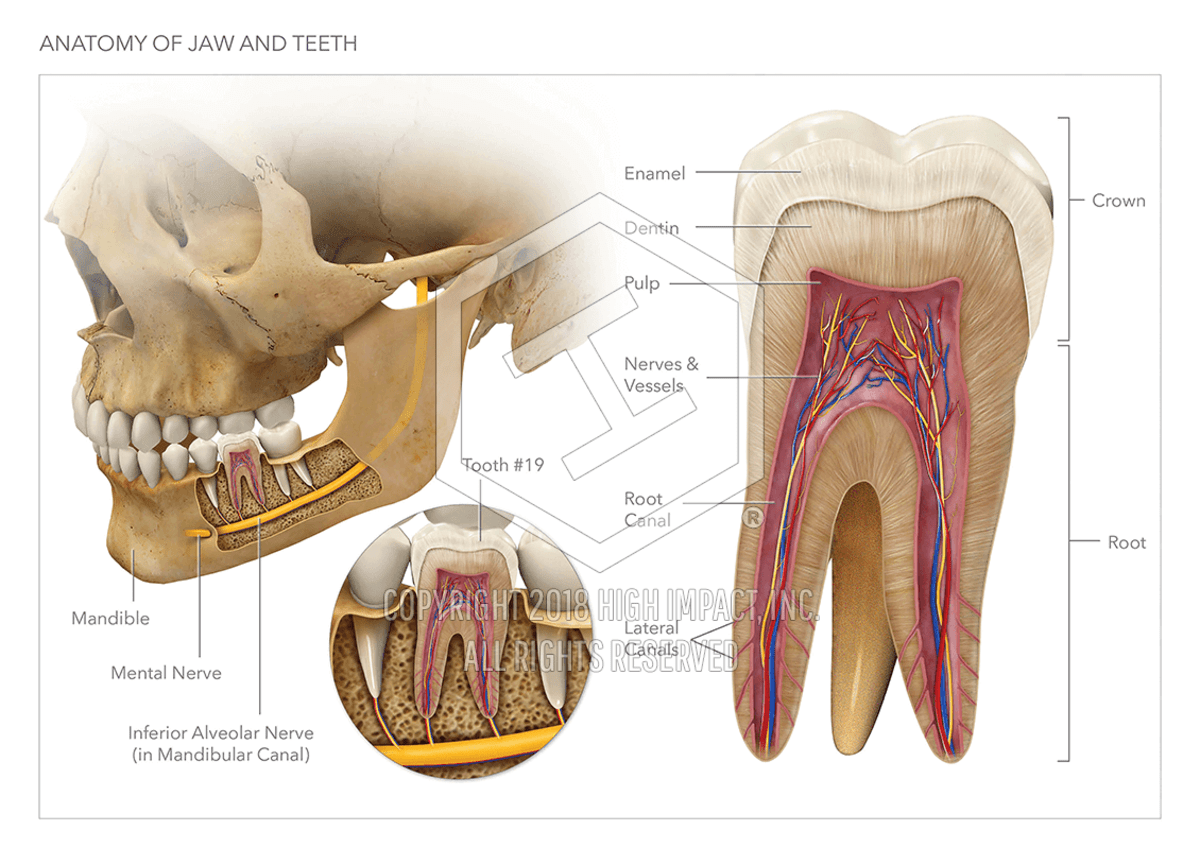 Anatomy Of The Jaw And Teeth High Impact Visual
 Muscles Of The Head And Neck Anatomy Pictures And Information
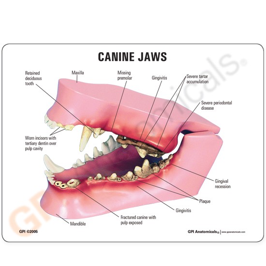 Canine Jaw Teeth Anatomical Model Lfa 9195
 Best Tmjd Physical Therapy Tmj Physical Therapists
 Anatomy Of Healthy Teeth And Dental Implant In Jaw Bone 3d Model
 Classic Human Skull Model With Opened Lower Jaw 3 Part
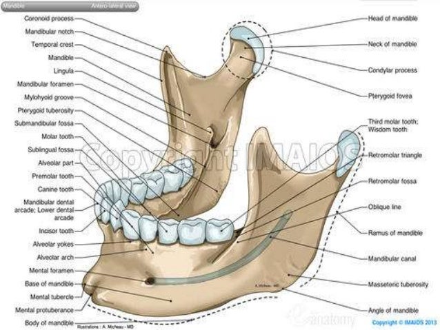 Anatomy Of Maxilla And Mandible
 Normal Anatomy Of The Jaw This Lateral View Of The Skull
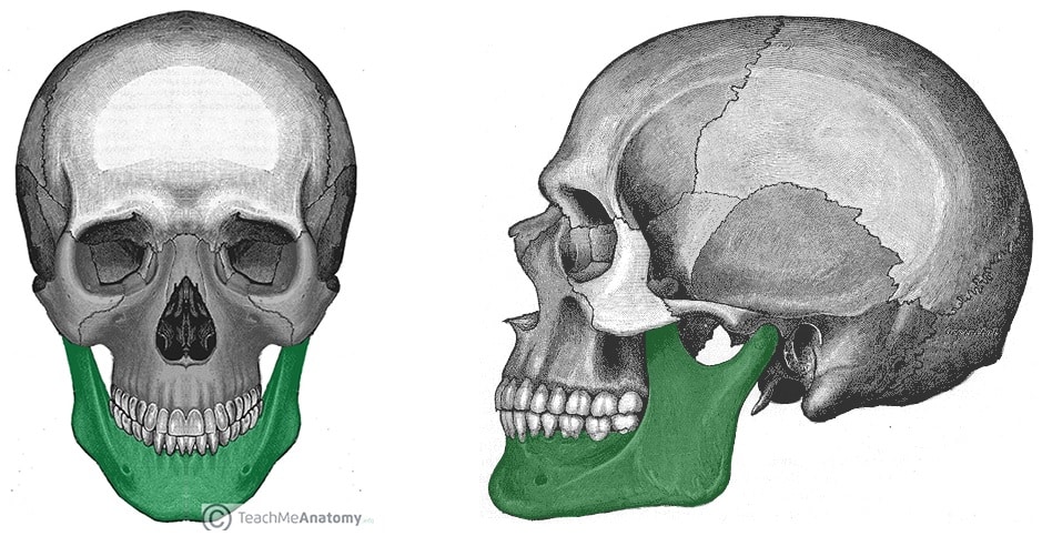 The Mandible Structure Attachments Fractures
Anatomy Stock Images Head Skull Jaw Side Skin
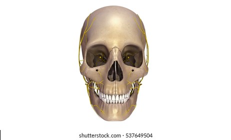 Royalty Free Human Jaw Bone Stock Images Photos Vectors
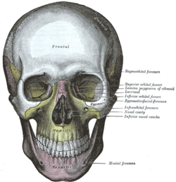
 The Skull Anatomy And Physiology
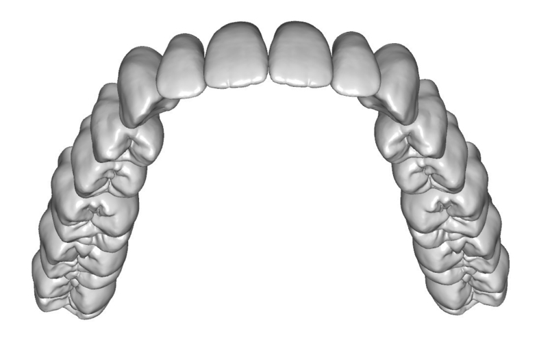 Teeth Anatomy And Morphology Upper And Lower Jaw


Posting Komentar
Posting Komentar