The cranial and facial bones and structures. The vagus nerve is responsible for heart rate gastrointestinal peristalsis and sweating to name.

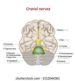
The first two nerves olfactory and optic arise from the cerebrum whereas the remaining ten emerge from the brain stem.

Cranial anatomy. Key points the vagus nerve cranial nerve x sends information about the bodys organs to the brain. The brainstem can be divided into three levels the midbrain the pons and the medulla. Cranial nerves have paths within and outside the skull.
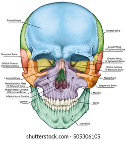
Detailed anatomy of the human skull. The brain is in charge of movement memory preservation and involuntary functions in the body. The cranial and facial bones and structures.
Examination of the cranial nerves allows one to view the brainstem all the way from its rostral to caudal extent. The skull of a normal bird usually weighs about 1 of the birds total bodyweight. The cranium skull is the skeletal structure of the head that supports the face and protects the brain.
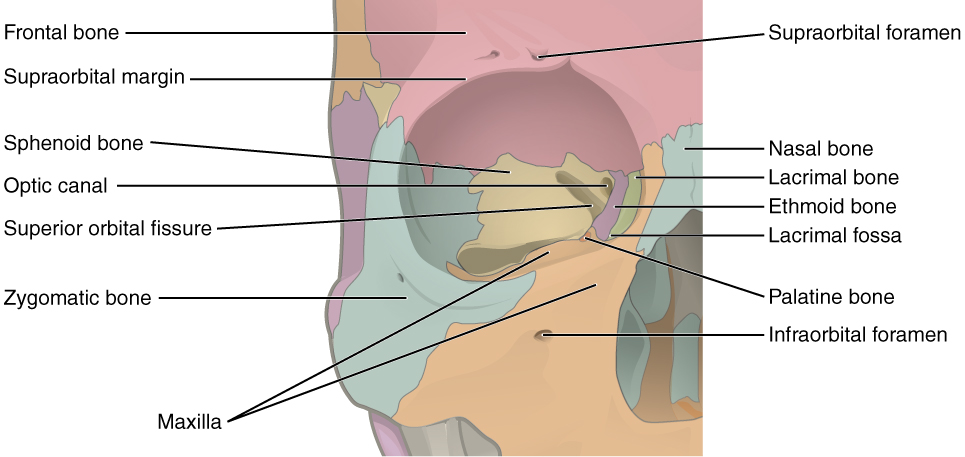
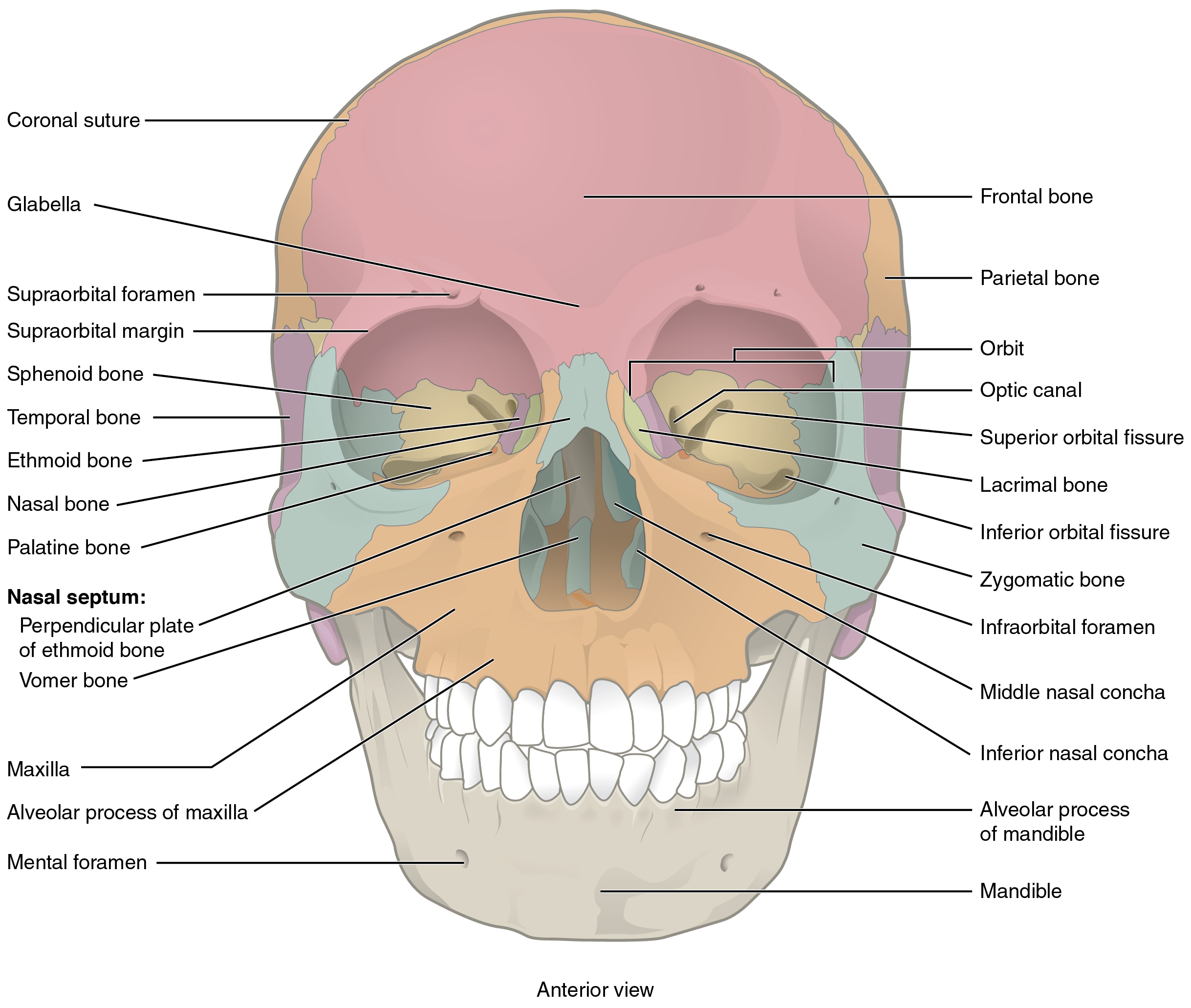
The facial bones underlie the facial structures form the nasal cavity enclose the eyeballs and support the teeth of the upper and lower jaws. The brain is in charge of movement memory preservation and involuntary functions in the body. There are many holes in the skull called foramina by which the nerves can exit the skull.
The cranial nerves are a set of 12 paired nerves that arise directly from the brain. The frontal top of head parietal back of head premaxillary and nasal top beak and the mandible bottom beak. These bones are important as they provide an articulation point for the 1st cervical vertebra atlas as well as the facial bones and the mandible jaw bone.
It is subdivided into the facial bones and the brain case or cranial vault link. The skull consists of five major bones. Cranial anatomy the brain is the control center of the nervous system and consequently the human body.
Cranial anatomy the brain is the control center of the nervous system and consequently the human body. The vagus nerve has axons that originate from or enter the dorsal nucleus of the vagus nerve. Comprised of six bones the frontal sphenoid ethmoid occipital parietal and temporal bones.
The paths within the skull are called intracranial and the paths outside the skull are called extracranial.
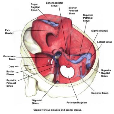
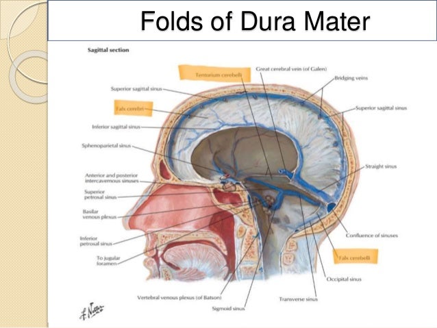 Cranial Cavity Department Of Anatomy
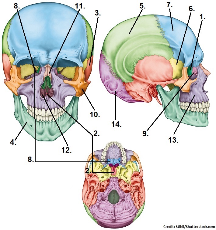 Skull Bones Quiz Cranial And Facial Bones

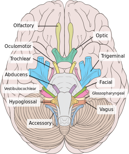
![]() Mixed Cranial Nerves Anatomy Course Fibers Functions
Mixed Cranial Nerves Anatomy Course Fibers Functions
 Cranial Anatomy Of Rhinolophus Euryale A 3d Mct Inferior
 4d Vision Human Anatomy Cranial Skull Model

 Cranial Nerve Anatomy Cranial Nerves Iowa Head And Neck
 Cranial Anatomy Of M Wachtleri Pzo 628 Based On Personal
Plos One Cranial Anatomy And Palaeoneurology Of The
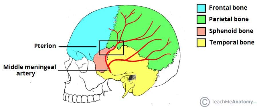 Bones Of The Skull Structure Fractures Teachmeanatomy
 1000 Cranium Stock Images Photos Vectors Shutterstock
 Cranial Anatomy Stock Vectors Images Vector Art
 Evo Devo Of A Grad Student Cranial Anatomy For Anth 2030
 Neurosurgery Operative Cranial Neurosurgical Anatomy
 The Skull Boundless Anatomy And Physiology
 Skull Base Anatomy Overview Anterior Skull Base Middle
 Cranial Nerves Wikipedia
Plos One Cranial Anatomy And Palaeoneurology Of The
 7 2 The Skull Anatomy And Physiology
 Cranial Nerves List And Functions Preview Human Anatomy Kenhub
 Brain Anatomy Goodman Campbell Brain And Spine
 7 2 The Skull Anatomy And Physiology

 The Cranial Fossae Teachmeanatomy
 Normal Cranial Anatomy The Greater Bilby


Posting Komentar
Posting Komentar