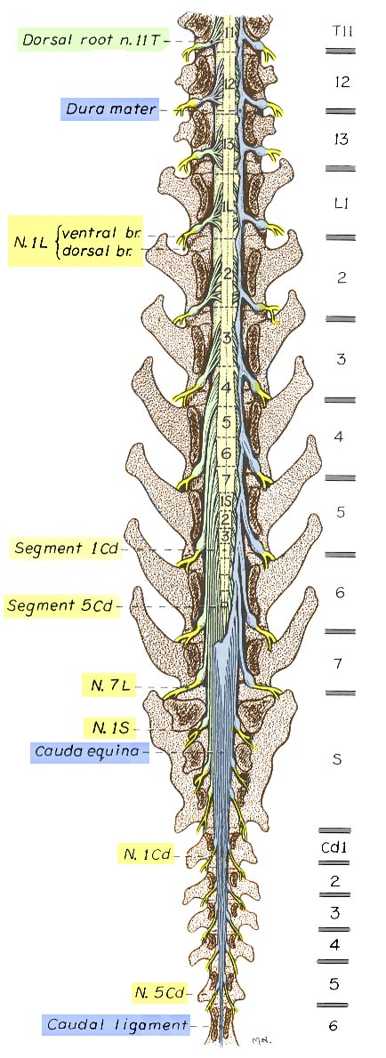
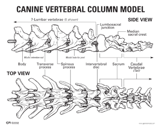
Normal dog have usually 7 lumbar vertebrae and 3 sacral vertebrae. Next comes the vertebra or spine.
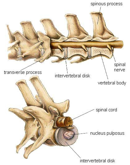
The spine is made up of block bones called vertebrae.

Spine anatomy dog. It is a long bone structure that encases the brain and contains a cavity called the orbit where the eye is located. It is elongated and extends to the end of the muzzle. One extremely important part of a dogs skeletal anatomy is the skull.
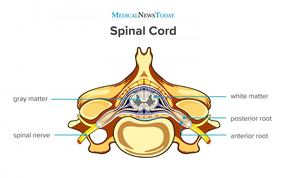
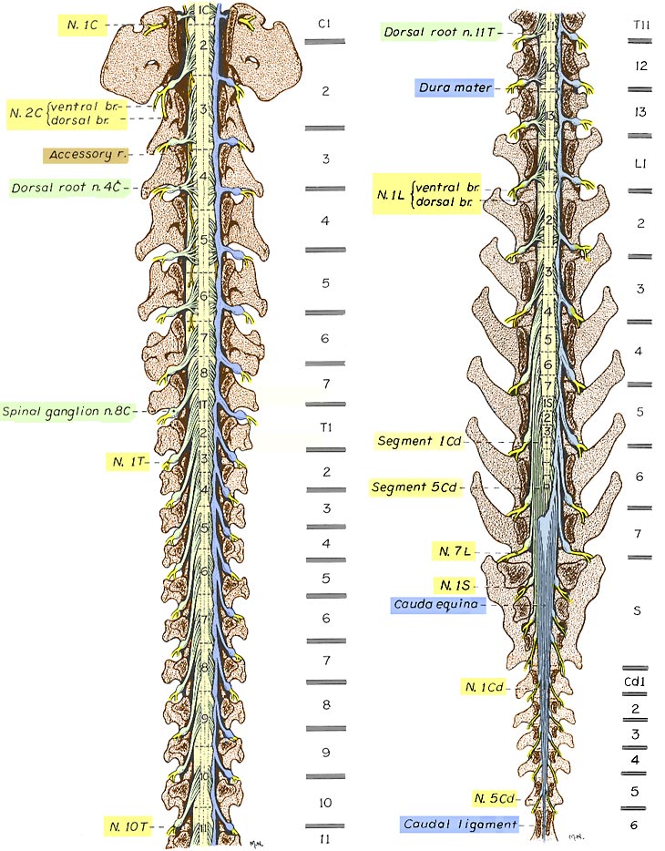
For educational purpose we decided to numerate lumbar vertebrae until 7 but if you have any question dont hesitate to contact us on imaios support. Each root is formed by rootlets which fan out along each segment of the spinal cord. In the dog there are 36 pairs of spinal nerves.
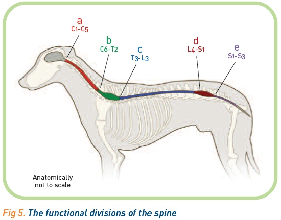
Dogs have a foot or paw at the end of each leg called the forefoot or hind foot depending on whether its front or back. Only one third is carried on their hind legs. In normal stance as shown in figure 5 2 a dogs spine is flexed at the atlantooccipital and atlantoaxial joints straight neither flexed nor extended in the remainder of the cervical spine extended at the cervicothoracic junction slightly lordotic in the thoracic spine and flexed or normally kyphotic in the lumbar spine.
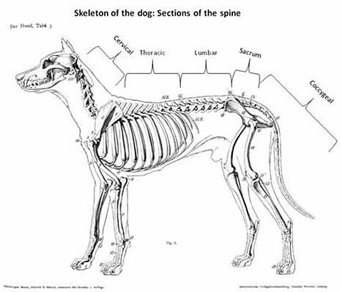
There is either a slightly flexed or extended sacrum on the lumbar spine depending on the tail posture. Dogs have seven cervical neck thirteen thoracic chest seven lumbar back three fused sacral tail bone and a variable number of tail vertebrae. The paw comes with nails sometimes called claws paw pads and usually dewclaws.
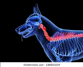
Dog leg anatomy just like humans have arms and legs dogs have forelegs and hind legs. It runs from the base of the head to the end of the tail. Spinal column anatomy physiology learning resources drag and drop dragster test your knowledge using drag and drop boxes canine spinal skeletal anatomy resources i ii iii canine spinal skeletal anatomy resources iv v canine whole spine skeletal anatomy resource.
Dog anatomy notice that the kidneys are not labeled on this picture. Image modified from hills pet nutrition atlas of veterinary clinical anatomy. Each is formed by a dorsal and a ventral root.
The dorsal root is formed by axons of afferent neurons sensory that have their bodies located at the spinal ganglion. Two thirds of a dogs body weight is carried on their front legs. A dogs spine is located along the top or dorsal side of a dogs body.
Unfortunately the dog used for this anatomical module presented a vertebral transitional anomaly with only 6 free lumbar vertebrae. The kidneys are tucked up close to the liver toward the spine.

 Small Animal Radiography Of The Scapula Shoulder Humerus
 Back Pain Elwood Vet
 Canine Whole Spine Skeletal Anatomy Resource Wikivet English
 Canine Anatomy Veterian Key

 Ivdd Surgery What If Your Vet Recommends Surgery For Your
 Anatomy Lumbar Dog Spinal Cord Nerve Anatomy Spinal
 Spine Anatomy
 Spinal Cord Anatomy Functions And Injuries
 Dog Bones Anatomy Dog Bones Anatomy
 Dog Spine Anatomy Images Stock Photos Vectors Shutterstock
 Comparative Anatomy Of The Horse Ox And Dog The Vertebral
 Spine Anatomy Video

 Pin By Peter Piggott On Health Nursing First Aid Dog
 Sitting With A Purpose Totofit
 Canine Vertebrae Model 9080 For Sale Anatomy Now
 French Bulldog Hemivertebrae Ufaw
 Vertebrae Thoracic
 Anatomy Of Domestic Dog And Cat Educational Chart Cool Wall Decor Art Print Poster 12x18
 Canine Anatomy Complete Set Of 3 Charts Buy The Set And Save
 Dachshund Anatomy Dachshund Dog Dachshund Puppies Weenie
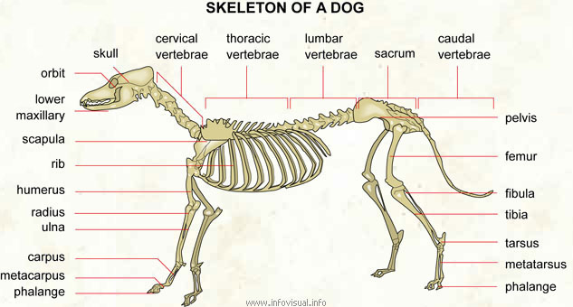 Skeleton Of A Dog Visual Dictionary
 Anatomy Pathophysiology Of Ivdd In Dogs Safari
 The Canine And Feline Vertebrae Veterian Key
 Lumbar Spine Of The Dog On Ct


Posting Komentar
Posting Komentar