The two sets anastomose frequently with each other. The deep veins accompany the arteries.
 Emergency Department Diagnosis Of Upper Extremity Deep
Veins of the upper extremity anatomy the upper extremity is equipped with both deep veins and superficial veins.

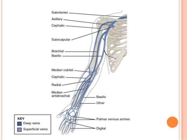
Upper extremity vein anatomy. Veins of the upper extremities are grouped into deep veins which are accompanying veins of arteries from which they derive their names latin. There is a vast tubular network of veins just below the skin of the upper extremity. The deep veins accompany the arteries and constitute the venæ comitantes of those vessels.
Vena comitantes and superficial veins. They are connected to the superficial system by perforating veins. The superficial veins starts on the back of the hand as a dorsal arch.
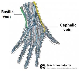
The drainage of blood allows for oxygenated blood to flow into the upper extremity. In green you can see arising laterally from the dorsal venous network is the cephalic vein and from the medial aspect of the dorsal venous network weve got the basillic vein which ive highlighted in purple. The deeper veins are buried well beneath the skin surface and run parallel to the arteries.
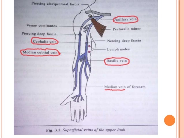
The superficial veins are placed immediately beneath the integument between the two layers of superficial fascia. The veins in the upper extremity drain the deoxygenated blood. The lymphatics of the upper extremity follows closely with the veins.
The superficial venous system of the upper limb essentially consists of two main veins which arise from the dorsal venous network. The lymphatics drainage start at the hand and drains toward the heart. The venous drainage of the upper limb is composed of superficial and deep vessels.
The veins of the upper extremity are divided into two sets superficial and deep. The veins do not perfuse the upper extremity with blood.
 Venous Lymphatic Drainage Of Upper Limb
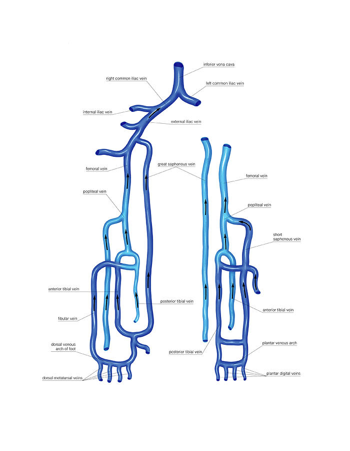
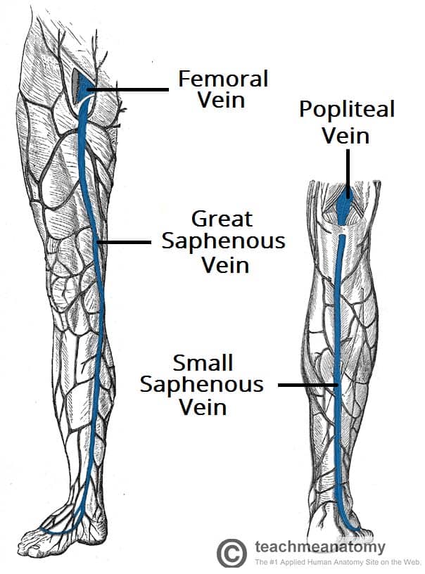 Venous Drainage Of The Lower Limb Teachmeanatomy

 Ultrasonography For Deep Venous Thrombosis Radiology Key
 Anatomy I Upper Extremity Deep Venous Return Diagram Quizlet
 Anatomy Revision Of The Upper Limb Lower Limb Back
 Superficial And Deep Veins Of The Upper Limb Diagram Quizlet
 Vein Wikipedia
 Cephalic Vein Wikipedia
 Basilic Vein Wikipedia
Upper Extremity Venous Thrombosis Management And Anatomy
 20 5 Circulatory Pathways Anatomy And Physiology
 Venous System Of The Upper Limb

 Venous Lymphatic Drainage Of Upper Limb
 Dentistry And Medicine Blood Supply Venous Drainage
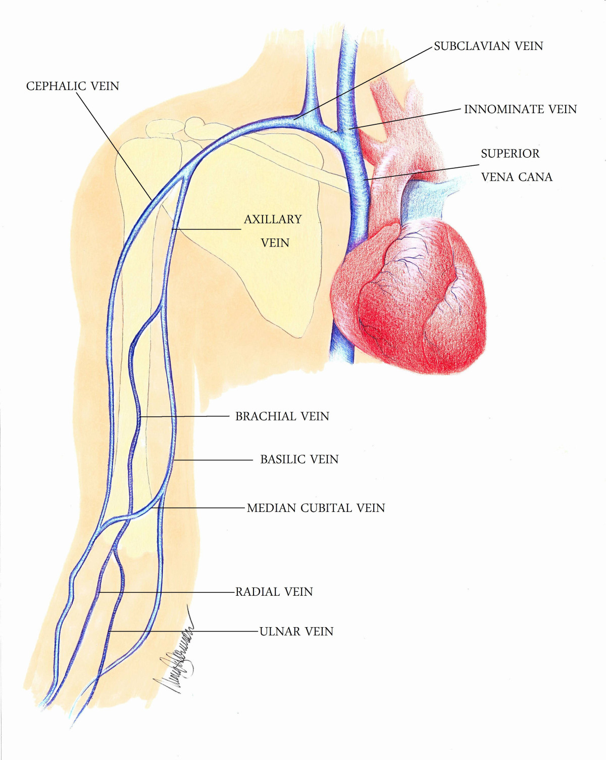
 The Veins Of The Upper Extremity And Thorax Human Anatomy
 Upper Extremity Venous Doppler Sonographic Tendencies
 Presentation1 Pptx Radiological Vascular Anatomy Of The
Presentation1 Pptx Radiological Vascular Anatomy Of The
 Figure 1 From Upper Extremity Deep Venous Thrombosis A
 Venous Drainage Of The Upper Limb Basilic Cephalic
![]() Veins Of The Upper Limb Anatomy Kenhub
Veins Of The Upper Limb Anatomy Kenhub
 6 Upper Extremity Locations For Autologous Avf Creation 1
![]() Veins Of The Upper Limb Anatomy Kenhub
Veins Of The Upper Limb Anatomy Kenhub


Posting Komentar
Posting Komentar