It can be subdivided further into four triangles which are detailed later on in this chapter. Veins external and internal jugular veins.
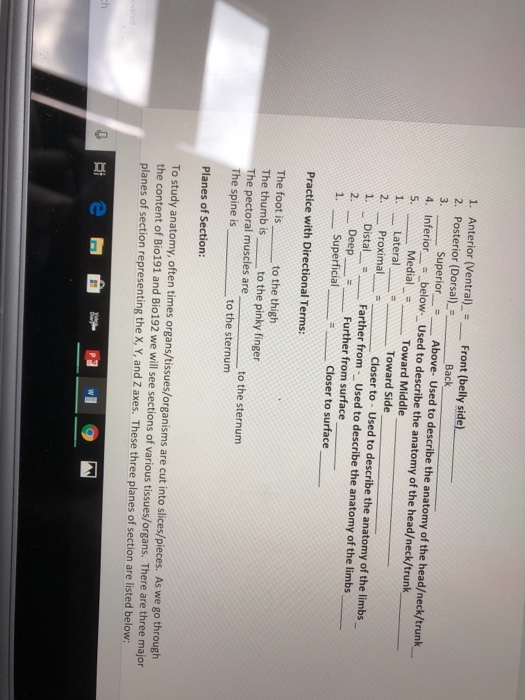
Neck anatomy nerves picture there are 8 spinal nerves that originate from the cervical spine.

Front of neck anatomy. The video has been produced by the das guidelines implementation group. Front of neck access training video das is pleased to share this video which demonstrates the recommended technique for surgical cricothyroidotomy. Other miscellaneous causes of front neck pain include sinusitis and throat infections such as tonsillitis.
Bone spurs in the neck although not common may be another cause of frontal neck pain. Medially sagittal line down the midline of the neck. These affect movements and sensation and some special organs such as hearing of parts of the head and neck.
The neck is the connection between the head and the body and is a very complex anatomic region. In the front of the neck the platysma muscle extends up from the chest goes over the collarbone and ends at the jaw. Muscle strain and herniated discs may also produce localized neck pain.
Investing fascia covers the roof of the triangle while visceral fascia covers the floor. Arteries common carotid arteries external internal carotid arteries. Superiorly inferior border of the mandible jawbone.
The spinal column contains about two dozen inter connected oddly shaped bony segments called vertebrae. They are the smallest and uppermost vertebrae in the body. The anterior triangle is situated at the front of the neck.
The majority of these nerves control the functions of the upper extremities and allow you to feel your arms shoulder and back of your head. In the front the neck goes from the bottom part of the mandible lower jaw to the bones of the upper chest and shoulder including the sternum and collar bones. It pulls down the lower face and mouth and causes wrinkles in these spots.
These injuries to the neck may be an acute condition or occur gradually over time. Movements of the neck includes. Flexion extension nodding yes and rotation shaking head no.
Therefore the most significant organs or structures at the front of the neck include. Laterally anterior border of the sternocleidomastoid. The infrahyoid muscles are the sternohyoid sternothyroid thyrohyoid and omohyoid.
The suprahyoid muscles are the digastrics stylohyoid mylohyoid and geniohyoid. Twelve pairs of cranial nerves emerge from the brain. The neck contains seven of these known as the cervical vertebrae.
In the back of the neck there is mainly muscle and of course the spine. Muscles platysma sternoclediomastoid supra and infrahyoid muscles groups of 4 muscles each. Muscles in the front of the neck are the suprahyoid and infrahyoid muscles and the anterior vertebral muscles see the images below.
The neck is the start of the spinal column and spinal cord.
 Anatomy Breathing Health Muscles Organ Systems Practice
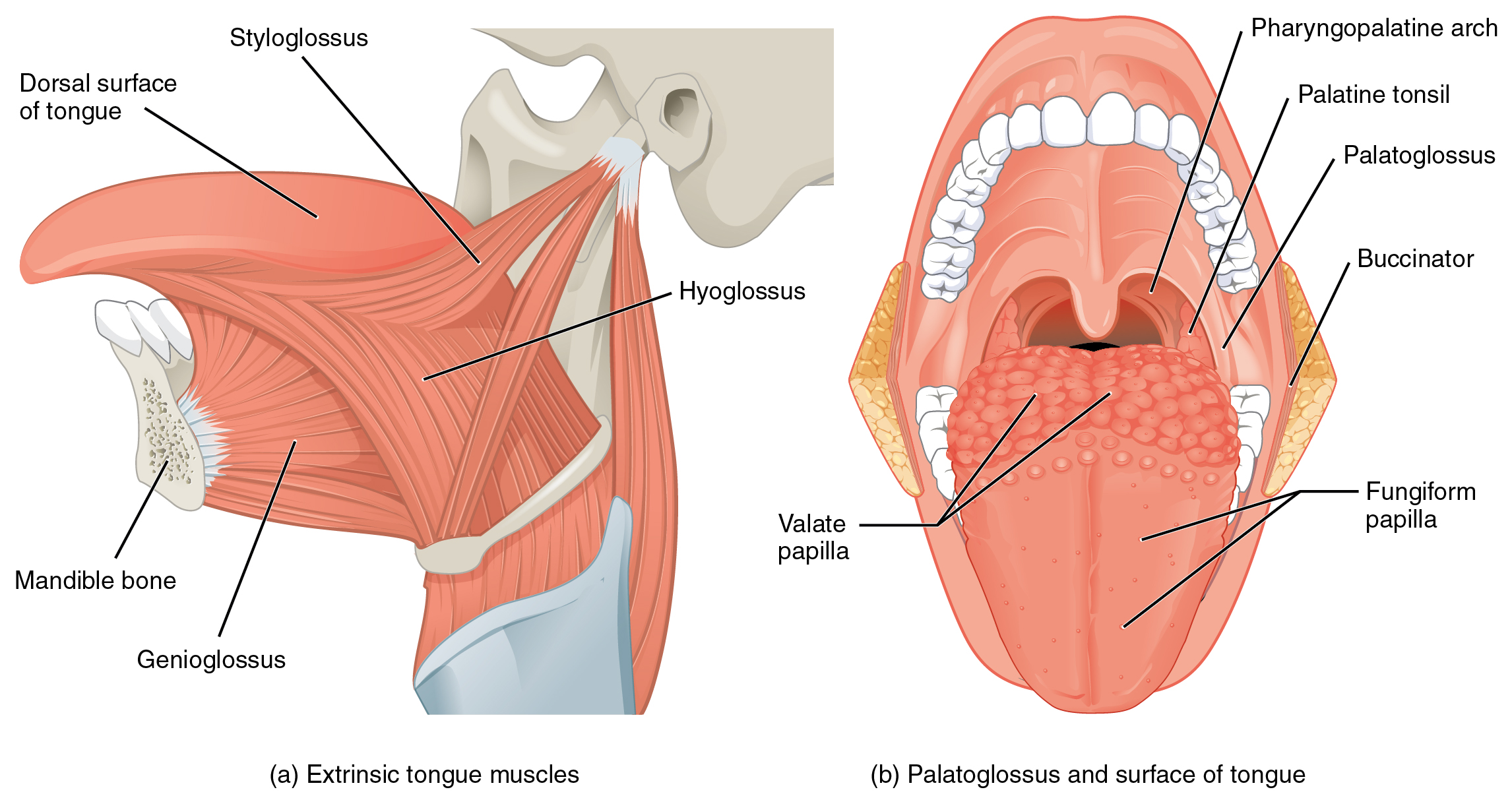 11 3 Axial Muscles Of The Head Neck And Back Anatomy And
 The Human Body Diagram Of Throat Wiring Diagram Images Gallery
 Ucsd S Practical Guide To Clinical Medicine

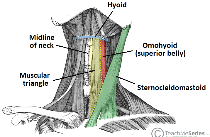 Anterior Triangle Of The Neck Subdivisions Teachmeanatomy
 Muscles Front Shoulder And Neck Diagram Quizlet
 Lymph Nodes Front Of Neck Stock Photos Page 1 Masterfile
 Oral Cancer Mouth Cancer Anatomy Headandneckcancerguide Org
 Face Neck Dissection Male Stock Image Image Of Neck
 Human Muscle Anatomy 3d Render On White Front Stock
 Endocrine Surgery Thyroid Cancer
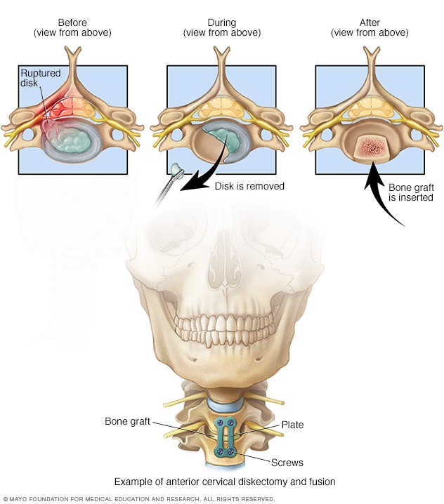 Fusion From Front Of Neck Mayo Clinic
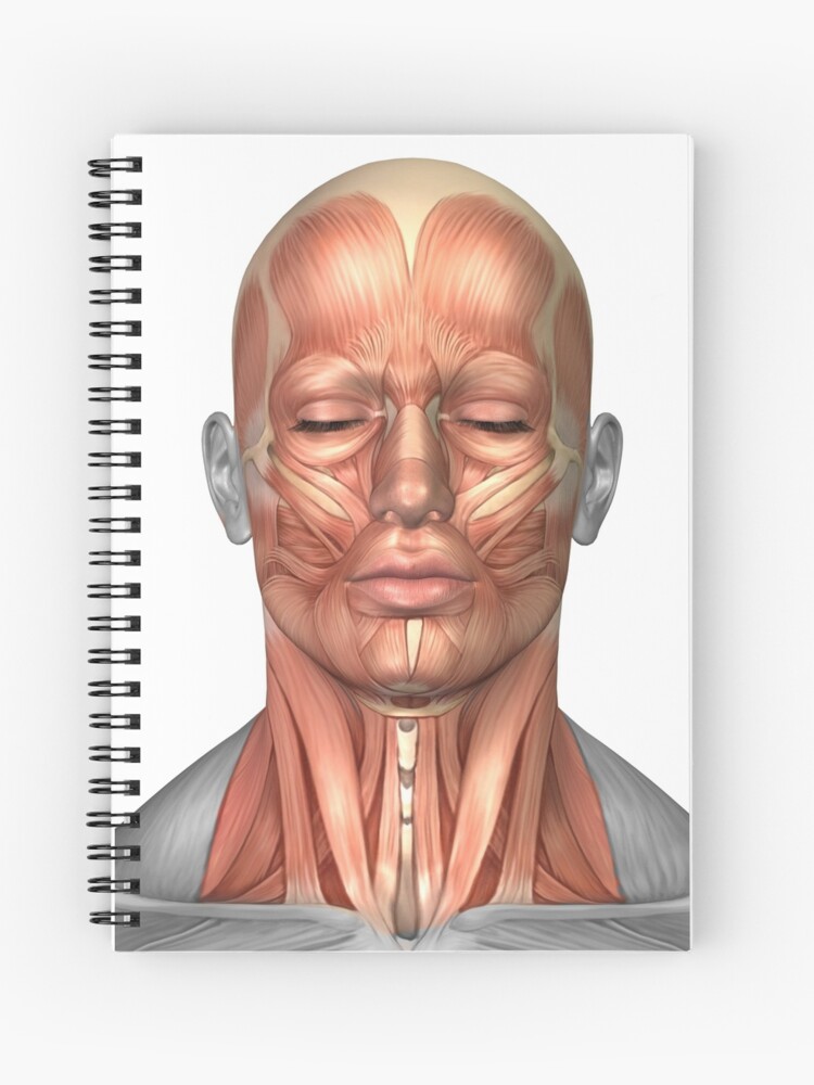 Anatomy Of Human Face And Neck Muscles Front View Spiral Notebook
Thyroid And Parathyroid Gland Anatomy Image Details Nci
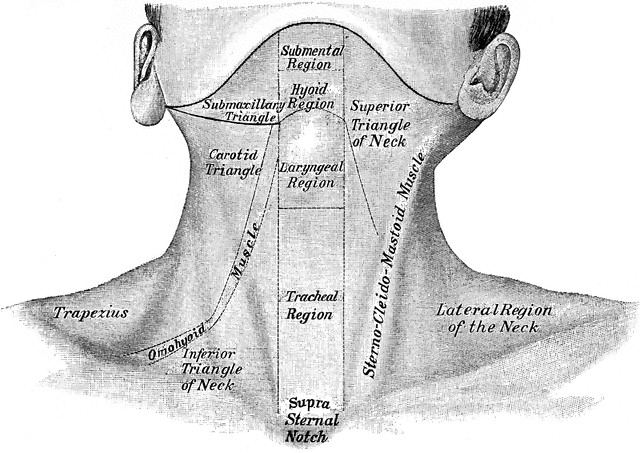 Front View Of Neck Clipart Etc
 Female Muscular Anatomy Semi Transparent Front View Clipart
 Neck Muscles Stock Illustration Illustration Of Drawing
 Introduction To Anatomy And Physiology Online Student
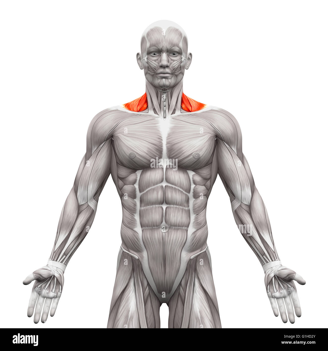 Trapezius Front Neck Muscles Anatomy Muscles Isolated On
Wetcanvas Artsschool Online Basic Anatomy L3
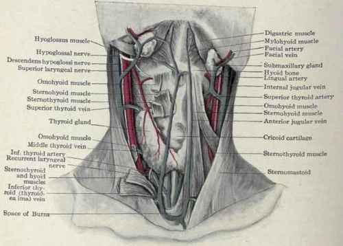

Posting Komentar
Posting Komentar