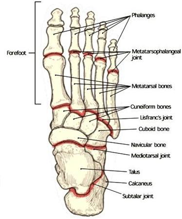
The forefoot contains the five toes phalanges and the five longer bones metatarsals. The foot is comprised of many bones joints tendons and ligaments including the plantar fascia and the achilles tendon.
 Muscles Of The Foot Part 1 3d Anatomy Tutorial
The foot contains 26 bones 33 joints and over 100 tendons muscles and ligaments.
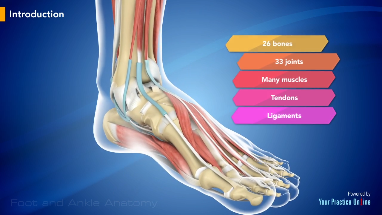
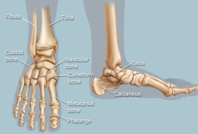
The anatomy of a foot. The feet are divided into three sections. The bones of the foot are organized into rows named tarsal bones metatarsal bones and phalanges. This may sound like overkill for a flat structure that supports your weight but you may not realize how much work your foot does.
Picture of foot anatomy detail. The foot is an extremely complex anatomic structure made up of 26 bones and 33 joints that must work together with 19 muscles and 107 ligaments to execute highly precise movements. This is the very front part of the foot including the toes or phalanges.
The foot is responsible for balancing the bodys weight on two legs a feat which modern roboticists are still trying to replicate. The foot contains a lot of moving parts 26 bones 33 joints and over 100 ligaments. The anatomy of the foot.
The foot is divided into three sections the forefoot the midfoot and the hindfoot. There are 14 toe bones two per big toe and three per each of the other four plus five metatarsals. The other bones of the foot that create the ankle and connecting bones include.
At the same time the foot must be strong to support more than 100000 pounds of pressure for every mile walked. This consists of five long metatarsal bones and five shorter bones that form the toes phalanges. The feet are flexible structures of bones joints muscles and soft tissues that let us stand upright and perform activities like walking running and jumping.
Learn about the anatomy of the foot. Soft tissues of the foot and ankle the soft tissues of the foot and ankle include ligaments tendons and fascia. These make up the toes and broad section of the feet.
Some of these structures were briefly discussed before. Picture of the feet. The first metatarsal bone is the shortest and thickest and plays an important role during propulsion forward movement.
 Foot Anatomy And Biomechanics Foot Ankle Orthobullets
 Facts About Feet Anatomy Snippets Complete Anatomy
 Deformities Of The Feet Anatomical Chart
:background_color(FFFFFF):format(jpeg)/images/library/11040/537_m_lumbricales_fkt.png) Ankle And Foot Anatomy Bones Joints Muscles Kenhub
Ankle And Foot Anatomy Bones Joints Muscles Kenhub
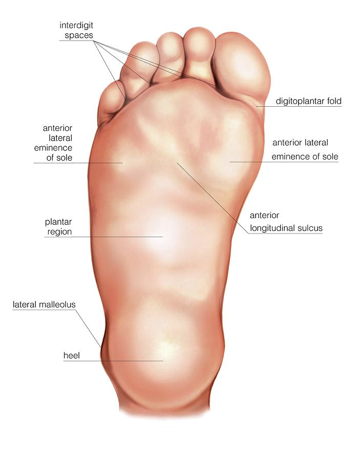 Anatomy Regions Of The Right Foot
 Regional Anatomy Foot At Texas Woman S University Studyblue
 Feet Human Anatomy Bones Tendons Ligaments And More
 Muscular Function And Anatomy Of The Lower Leg And Foot
 The Foot Advanced Anatomy 2nd Ed
 Understanding And Caring For Your Feet Breaking Muscle
Human Being Anatomy Skeleton Foot Image Visual
 Anatomy Of The Foot Comprehensive Orthopaedics
Anatomy Of The Foot And Ankle Bergen Family Foot Care
 Anatomy Of The Foot North Arkansas Podiatry
 Anatomy Of The Foot And Ankle With Foot Drop Deformity
:watermark(/images/watermark_5000_10percent.png,0,0,0):watermark(/images/logo_url.png,-10,-10,0):format(jpeg)/images/atlas_overview_image/939/S4mPs96snOyWo6sqWiPJ9g_muscles-foot_english.jpg) Diagram Pictures Muscles Of The Foot Anatomy Kenhub
Diagram Pictures Muscles Of The Foot Anatomy Kenhub
 Anatomy Specific Bones Of The Feet
 Us 24 99 Medical Anatomy Human Foot Normal Foot Flat And Arched Foot Anatomy Model Human Sketelon Model Flat Feet High Arched Feet Model In
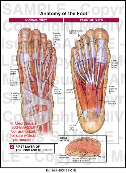 Anatomy Of The Foot What Is Special About It
 Illustrated Anatomy Of The Foot A The Cuneiforms Cuboid
 Foot Anatomy Foot And Ankle Bones Ligaments Tendons And
 Foot And Ankle Anatomy Allen Tx Foot Doctor
 Anatomy Of The Foot Medical Art Library
Foot Anatomy Foot Ankle Lower Leg Orthopedic Assessment
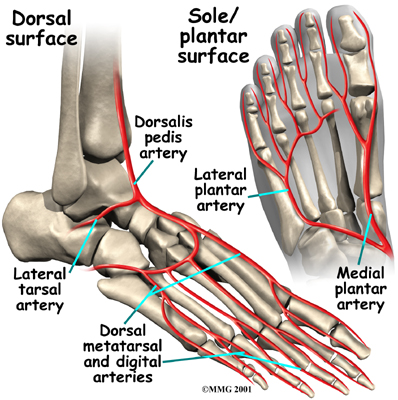 Physical Therapy In Columbia For Foot Anatomy
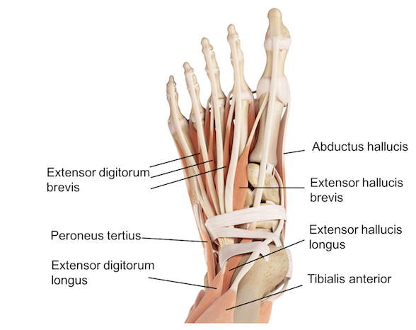
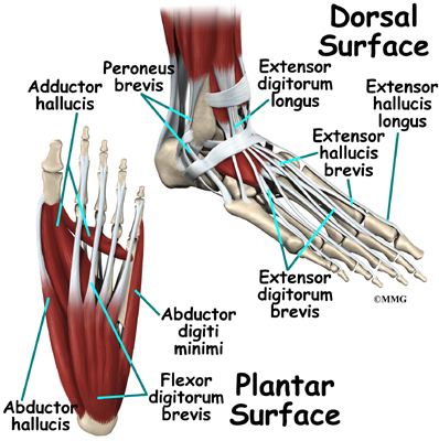


Posting Komentar
Posting Komentar